Case report
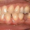
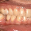
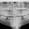
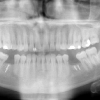
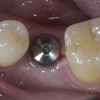
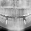
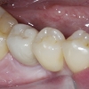
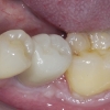
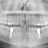
This case report involved a multi-disciplinary approach to properly replace missing lower first molars in this patient. Antonio presented with chief complaint of missing #19 and 30 which were extracted many years ago. He reported some difficulty eating and wanted to replace them. After, a comprehensive dental evaluation by his dentist, Dr. Giannini, patient was referred to Dr. Gerlein for orthodontic evaluation and Dr. Kazemi for dental implants. Examination revealed mesially inclined second molars and missing #19 and 30 with significant horizontal atrophy. It was clear that predictable replacement of his missing teeth required first creating adequate space orthodontically, followed by site augmentation for implant supported crowns.
Treatment
1. Following phase 1 dentistry, patient was seen by Dr. Gerlein to begin orthodontic treatment to upright second and third molars.
2. Patient was recommended to have his third molars removed to simplify uprighting and create more space for hygiene, but he did not want to lose them.
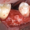
3. Upon completion of the orthodontic phase, the first surgical phase consisting of bone augmentation was performed. This was accomplished with an onlay graft in combination with a particulate graft and GTR membrane to enhance horizontal bone. Bone was harvested from the ramus area.
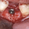
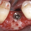
4. Six months later, implants #19 and 30 were placed in single stage fashion using a surgical guide prepared from a complete wax up.
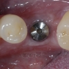
5. Following 3 months of healing, the implants were checked and prepared for the prosthetic stage by Dr. Giannini.
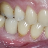
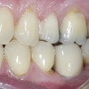
6. Implant level impressions were obtained and ceramic abutments and crowns were fabricated and placed.
Limiting factors
We had recommended the patient to have the third molars removed to allow more distalization of the second molar but he did not want to lose his third molars. There were some limitations on how much uprighting could be accomplished due to third molars. Patient was informed of the implications (less space in the first molar sites and smaller teeth) and he accepted. Even though second molars remained in less ideal axial position, the implant restorations are in great function and accessible for hygiene and the tissues remain healthy. Ideally, the third molars in such case should be extracted to allow the orthodontist recreate a more ideal space for the first molars.
Success factors
1. The diagnostic work up began with the end in mind.
2. Orthodontic treatment to create adequate space for first molars.
3. Site development with bone grafting. This allowed proper size implants with excellent tissue topography and emergence.
4. A proper surgical guide allowed accurate placement of the implants.
5. The surgeon , orthodontist, and restorative dentist working in collaboration, each doing what they do best.
