Case report
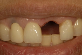
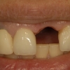
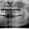
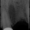
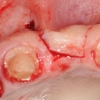
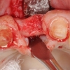
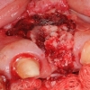
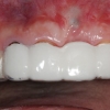
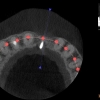
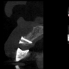
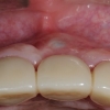
This patient presented to us for evaluation and treatment for replacement of tooth #9. Tooth #9 had been extracted a few months prior to her visit and the adjacent teeth had single crowns. The site of tooth #9 had clear deficiency of both bone and soft tissue. A CBCT demonstrated horizontal deficiency of bone. The treatment plan included bone and soft tissue augmentation and placement of a single dental implant to support a crown.
Treatment:
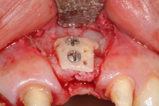
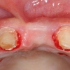
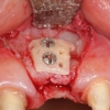
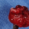
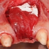
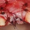
Dr. Keith Progebin, the treating prosthodontist, first fabricated a new provisional restoration using #8 and 10 as abutments. This provided some guidelines on the future treatments. Dr. Kazemi performed a bone graft augmentation using an onlay technique. Bone was harvested from the ramus and fixated with screws. Additional bone graft was complemented using particulate autogenous bone, mineralized freeze dried bone, and PDGF. A GTR membrane was placed and the incision was closed in tension-less fashion.
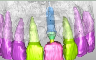
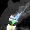
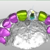
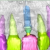
After a six months, a radiographic guide was prepared and used for the new CBCT. This allowed precise assessment of the underlying bone and its relationship to the intended implant and crown. Using the 3-D model, a surgical guide was fabricated translating the work-up to a guide that can be used during implant placement.
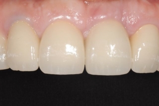
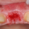
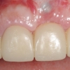
The implant was placed along with a connective tissue graft from the palate. A delayed provisional restoration was fabricated and maintained for about 3 months. This allowed proper design of the soft tissue architecture. Final crowns were then fabricated according to the new tissue form.
