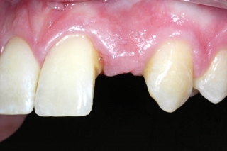
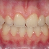
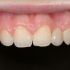
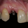
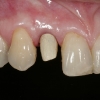
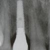
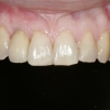
Replacement is congenitally missing lateral incisors has unique challenges that often requires a multi-disciplinary approach for success and predictable results.
Implant site development:
The missing lateral incisors may be first discovered in patients between 7 and 10 years of age, often presenting with retained maxillary primary lateral incisors. At this stage, if the crown of the permanent canine is apical to the primary canine root, it may be necessary to extract the primary lateral incisor to allow eruption of the permanent canine. If the permanent canine is allowed to erupt into the lateral incisor position, following orthodontic movement distally, an increased buccolingual alveolar width is established which seems to remain stable over time. In older patients, it is quite common that the lateral incisor site is atrophic and therefore site development will require bone and soft tissue grafting.
Space appropriation:
Four methods of determining appropriate spacing for missing lateral incisors:
- Golden proportion: Each tooth is about 61.8% wider than the tooth distal to it
- Measure contralateral incisor if it has a normal width.
- Bolton analysis
- Diagnostic wax-up (The most predictable method)- General width ranges from 5-7 mm
Requirements for implant placement:
- 1.5 to 2 mm space is recommended between head of implant and adjacent teeth, although this is often difficult to accomplish.
- The goal should be to achieve a minimum of 6 mm space.
- With a 7 mm wide space and use of a traditional 3.7 mm implant, an approximate 1.5 mm space is maintained between implant and adjacent teeth.
- With a 5 mm space, a 3mm diameter implant maybe used, leaving 1 mm on each side. The response of the soft tissue and papilla should be considered in this situation and approached with great caution.
Problems with inadequate space between root apices are generally due to improper mesiodistal root angulation. A periodic periapical x-ray can indicate the progress as orthodontist diverges the roots. In certain patients, it may be impossible to achieve acceptable space between roots even though the coronal spacing may be ideal. An example is patients with Class III tendency requiring proclination of the incisors.
Timing of implant placement
It is crucial the implant are placed once patient has completed facial growth. Placement of dental implants before completion of growth will result in poor alignment and aesthetic problems as implant remains in apical position. Also, the orthodontic treatment and site space preparation must be completed prior to placement of the dental implant.
Two predictable techniques to assess completion of growth:
- Serial cephalometic radiographs taken 6 months to one year apart
- Cervical vertebral X-ray- Described by Dr. McNamara- Click to see full article:
Cervical Vertebral Maturation (CVM)
Interim tooth replacement after orthodontics:
Until facial growth is complete and implants can be placed, options are:
- Removable retainer with a prosthetic tooth- Good for short period of time before implant can be placed.
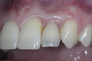
- Resin-bonded fixed partial dentures- More appropriate if implant is is planned in several years. (photos courtesy of Dr. Benjamin Watkins).
Implant site development with bone grafting:
Missing lateral incisors sites often present with horizontal atrophy, with inadequate foundation for implant placement and proper tissue emergence and aesthetics. Therefore bone and soft tissue grafting may be necessary to develop proper implant site. The interim prosthesis can provide good information with regard to amount of bone and soft tissue deficiency and the appropriate surgical approach for site development. Implants can be placed five to six months after bone augmentation surgery.
Implant placement
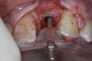
Precision implant placement requires a well fabricated surgical guide
A surgical guide from wax-up or CBCT is necessary to guide the surgeon in appropriate positioning of the dental implant. If implant is very stable, it is possible to place an immediate provisional restoration. Otherwise it is placed in delayed fashion following implant integration.
Implant provisionalization
An implant supported provisional restoration guides the soft tissue and creates ideal gingival architcture and cosmetic results.
An implant-supported provisional restoration is placed on day of exposure or immediately if possible. Soft tissue is allowed to mature for at least 3 months before final impression. During this period, soft tissue architecture can be designed and tooth is shaped for optimal aesthetics.
Final restoration
The final restoration is fabricated once the soft tissue architecture and tooth aesthetics has been developed to satisfaction. Decision between ceramic vs titanium abutments and ceramic vs. PFM crowns are made based on patient smile line, soft tissue thickness, aesthetic requirements, and patient expectations.
(Courtesy of Dr. Benjamin Watkins)
