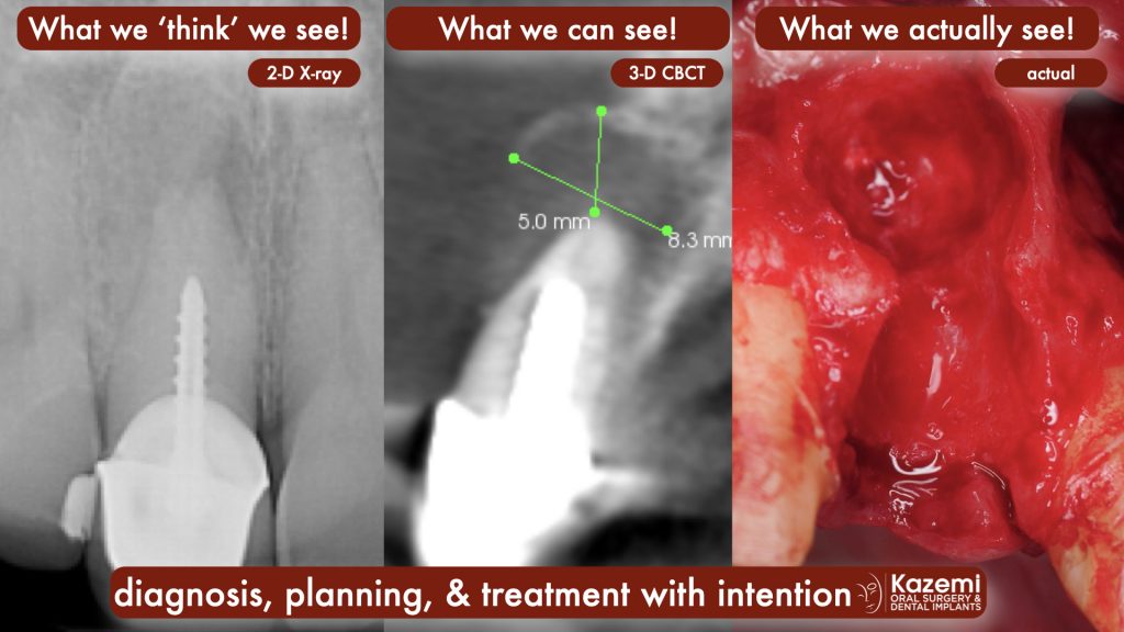
2-dimensional dental X-rays (peri-apical x-rays and panorex) were the only diagnostic imaging in dentistry until availability of 3-dimensional cone beam CT scan (CBCT) in early 2000. Currently 3-d imaging is not a routine or required X-ray in dentistry and hence many patients with conventional 2-D X-rays remain undiagnosed for common dental diseases such as abscess, cysts, and other forms of infections. Such conditions are not visible on 2-D x-rays until they have advanced and caused significant amount of decalcification and bone loss. And even then the extent of such conditions can not be accurately assessed without a 3-D cone beam CT scan. In an earlier article, ‘What X-rays Should Dental Patients Have’, we described a recommended criteria for X-rays depending on specific dental health and needs of individual patients.
When dental infection is suspected or noted on 2-D x-rays, clinicians must recommend a cone beam CT scan (CBCT) for proper diagnosis, planning, and treatment.
