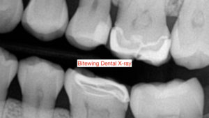
 Bitewing dental X-rays are commonly used at regular hygiene or check-up visits to detect dental caries. Unfortunately, they have limited field of view and do not show the entire roots and their surrounding structures. In a previous article in June 2019, “ What X-rays Should Dental Patients Have ”, we proposed a set of guidelines for type and frequency of dental X-rays based on specific patient needs and oral health. And in another article in August 2019, “ Why Cone Beam CT Scan (CBCT) is a Must for Diagnosis of Teeth-Related Infections “, we discussed the importance of CBCT (cone beam CT scan) in proper diagnosis of lesions often not evident in routine dental X-rays.
It is not uncommon for many patients to get only bitewing X-rays during their regular check-up visits. Whether it is to limit radiation or cost, bitewing X-rays are simply inadequate and do not provide proper diagnosis of pathological conditions which may remain asymptomatic and hence not discovered. Bitewing X-rays should be used in conjunction with other imaging studies, such as panorex, cone beam CT scan (CBCT), or even a full mouth series as relevant to the needs of individual patients. As demonstrated in two cases described below, sole use of bitewings will lead to missed diagnosis and compromised patient care:
Case #1:
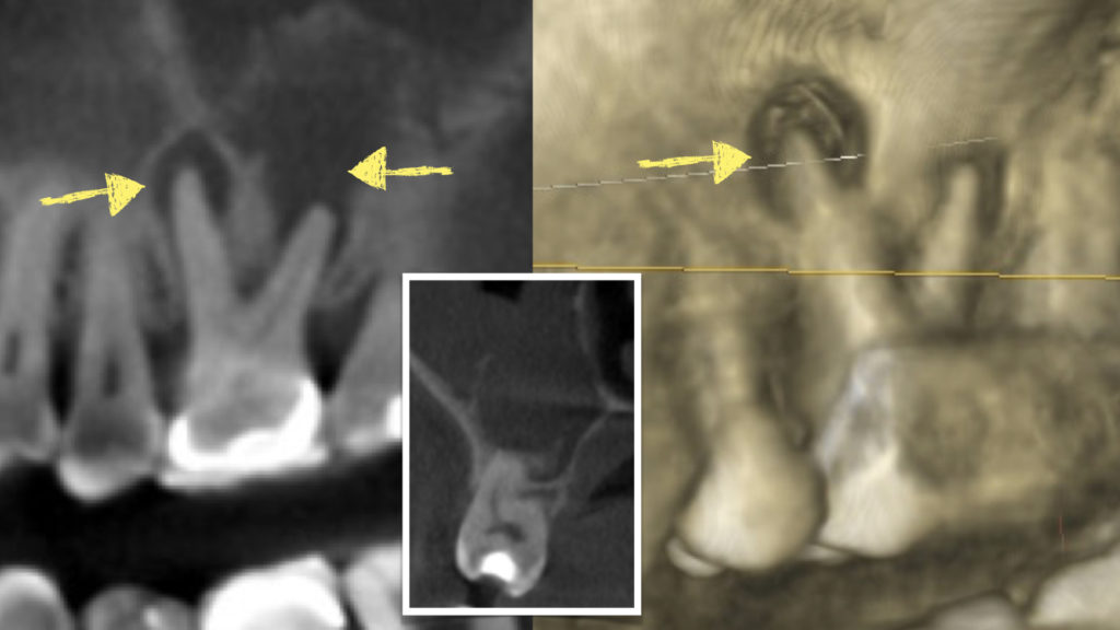
A 60-old patient was referred for evaluation of jaw pain following a fall. To rule out fractures, a cone beam CT scan was obtained. While there were no fractures, an upper left molar was incidentally noted with extensive loss of bone at the tip of the roots and lesions extending into the sinus cavity. Patient was completely asymptomatic and unaware of the condition. The cone beam CT scan showed large peri-apical lesions with perforation into the sinus and evidence of sinusitis. Patient said that he had seen his dentist regularly every 6 months and had only bitewing X-rays during the last 2 years. Endodontic and surgical treatment options were discussed. However, due to guarded prognosis and extensive erosion into the sinus cavity, patient was recommended for tooth extraction, excision of the infection and peri-apical cyst, repair of the perforation using layered grafting technique along with platelet rich fibrin. Earlier detection may have saved the tooth with only a non-surgical endodontic (root canal) treatment and a restoration.
Case #2:
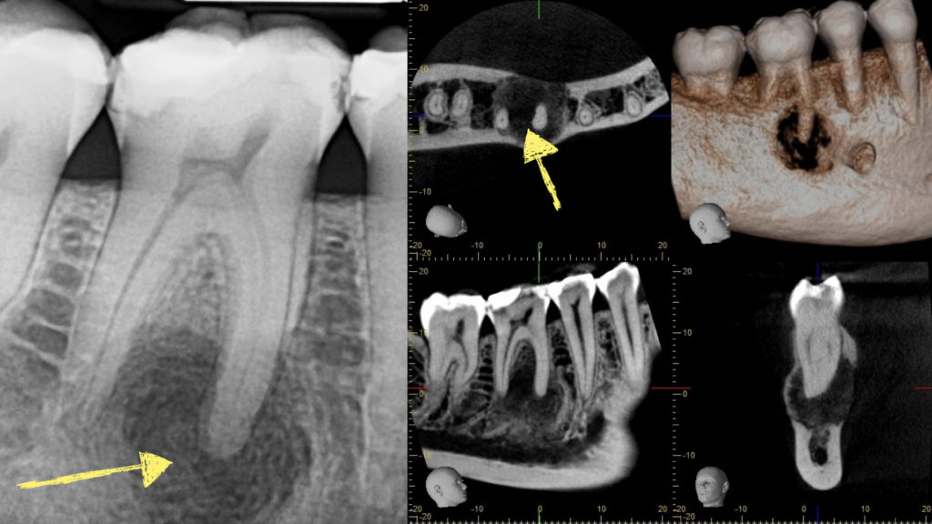
Another patient, a 35-year old female, came to us for evaluation of an upper tooth with gingival pain and bleeding. A panoramic X-ray was obtained to assess the overall teeth and the supporting jaw bone. The upper teeth appeared healthy and the gingival pain and bleeding was attributed to advanced gingivitis. However, during review of the panoramic X-ray, a large radioleucency (dark area) was noted involving the lower right first molar. Patient was completely asymptomatic and was not aware of any problems. She had been seeing her dentist every 6 months for check-up and cleaning and reported having only bitewing X-rays during the last 4 years. A CBCT was obtained which showed an extensive erosive lesion involving the entire width of the jaw bone with significant extension around the roots. An immediate biopsy was recommended to establish diagnosis and treatment plan. As in the patient described above, other types of X-rays would have led to earlier detection.
Both patients were quite surprised having such extensive lesions associated with their teeth even though they had seen their dentists continually and regularly for checkup and hygiene. Both asked “how was that missed?” Of course, with absence of any signs and symptoms, the dentists may not have seen a rationale to take other X-rays with larger field of view which would have certainly resulted in earlier detection.
This supports the rational for regular comprehensive X-rays at shorter intervals as such lesions can develop and expand quite rapidly resulting in extensive erosion and destruction of jaw bone and adjacent structures.
It is important that as dental professionals, we do not rely solely on bitewing X-rays and recommend other X-rays with expanded fields of view when appropriate. Likewise, it is critical for patients to discuss with their dentists the type and frequency of dental X-rays appropriate for them to assure a stable state of oral health.
Dr. H. Ryan Kazemi is a board-certified oral and maxillofacial surgeon in Bethesda, MD