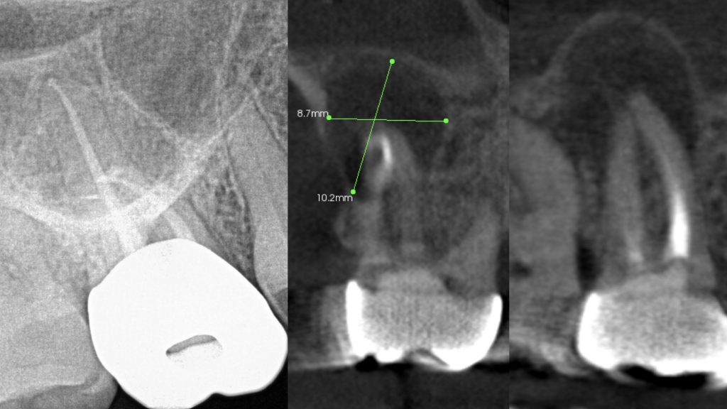
When patients present with tooth-related pain or swelling, the first line of radiograph is often a peri-apical X-ray or panorex which may or may not show abnormalities associated with an existing infection. It is well-recognized that periapical dental X-rays or panoramic X-rays are very limited in their ability to diagnose odontogenic infections or other forms of pathology. This is because such X-rays are flat and 2-dimensional. Here is an example of a patient who had received root canal therapy (endodontic treatment) on her upper molar several years ago and just recently developed pain in the region. A peri-apical X-ray was taken by her dentist which appeared normal.

With continued symptoms, patient’s endodontist immediately recommended a cone beam CT scan (CBCT) which provides a 3-dimensional view of the site. The scan showed a large area of infection characterized by bone loss measuring 10mm by 9 mm and approaching the maxillary sinus. With guarded prognosis, treatment consisting of tooth extraction, removal of the infection / cyst, and site bone grafting was immediately performed to prevent further expansion and possible perforation into the maxillary sinus.
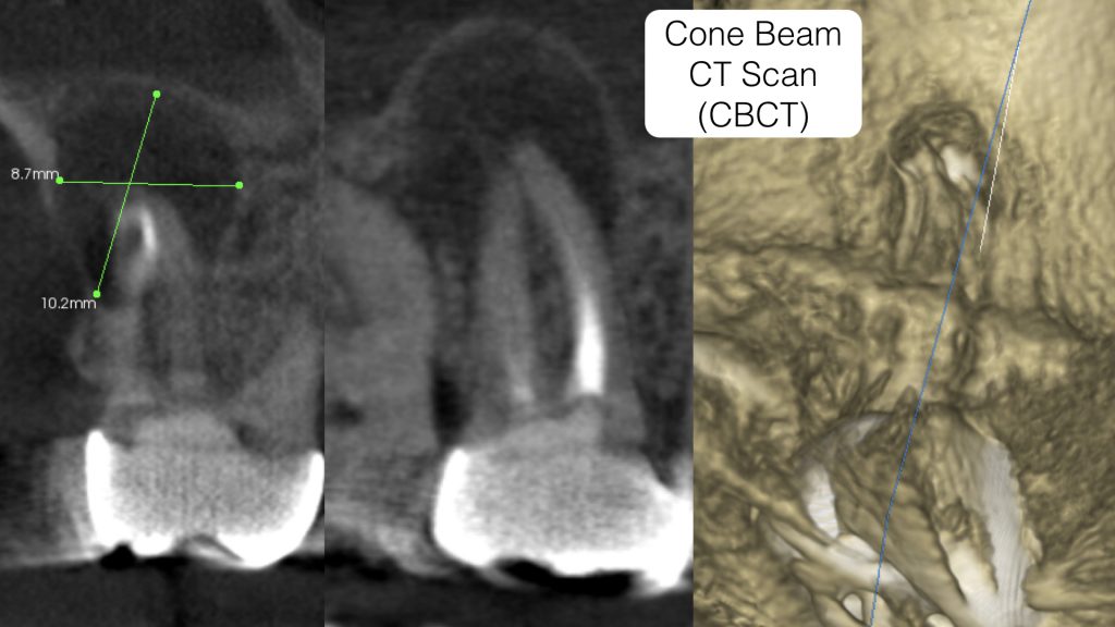
Peri-apical X-rays have limited benefits in diagnosis. When presented with symptoms, a cone beam CT scan (CBCT) is the only dependable radiographic technique for accurate and early diagnosis.
Dr. H. Ryan Kazemi is a board-certified oral and maxillofacial surgeon in Bethesda, MD