Pigmented oral lesions refer to any abnormal areas of discoloration or pigmentation in the oral cavity. These lesions can vary widely in appearance, ranging from benign conditions to potentially malignant or malignant lesions. Some common pigmented oral lesions include:
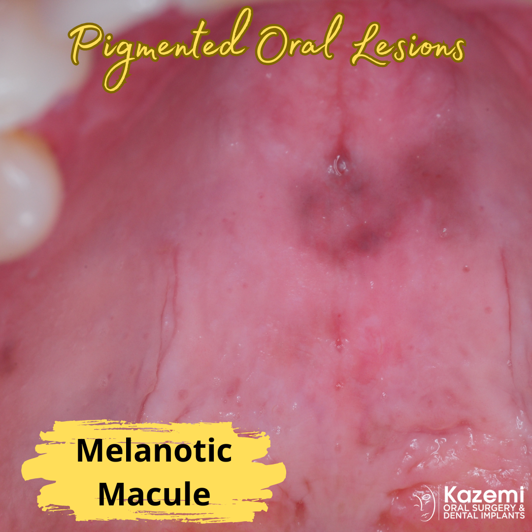
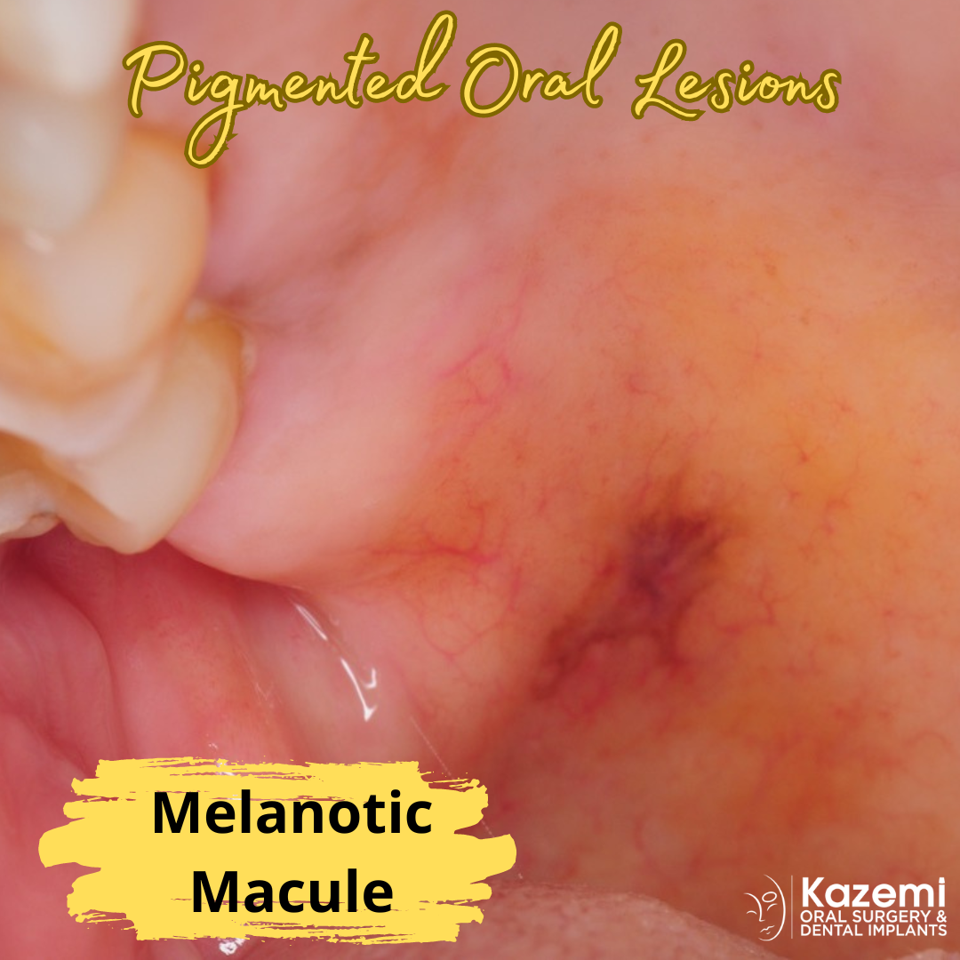
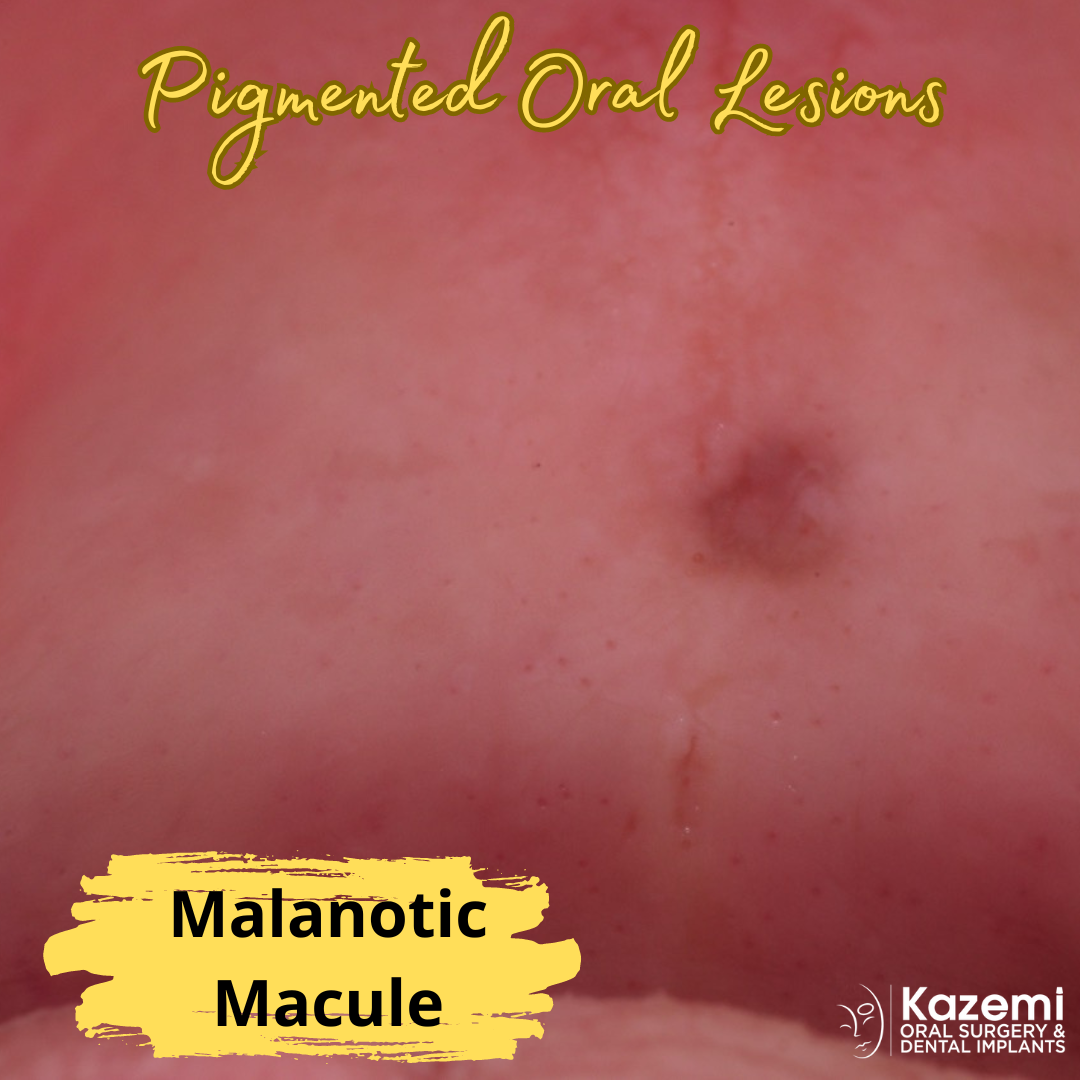
- Melanotic Macule: As described earlier, melanotic macule is a benign pigmented lesion typically found on the lips or oral mucosa.
- Oral Melanotic Nevi: Similar to melanotic macules, oral melanotic nevi are benign proliferations of melanocytes in the oral mucosa. They may appear as brown to black patches or spots and are usually asymptomatic.
- Oral Melanoma: Oral melanoma is a malignant tumor arising from melanocytes in the oral cavity. It can present as a pigmented lesion with irregular borders, asymmetry, and changes in color over time. Oral melanoma is rare but can be aggressive and potentially life-threatening.
- Amalgam Tattoo: Also known as focal argyrosis or focal argyria, is a benign condition that occurs when particles of dental amalgam become embedded in the oral tissues
- Oral Lichen Planus: Oral lichen planus is a chronic inflammatory condition that can affect the oral mucosa, causing various types of lesions, including pigmented ones. These lesions may appear as blue-gray or brown patches or lines.
- Smoker’s Melanosis: Smoker’s melanosis is a benign condition characterized by brown to black pigmentation of the oral mucosa, typically seen in individuals who smoke tobacco products.
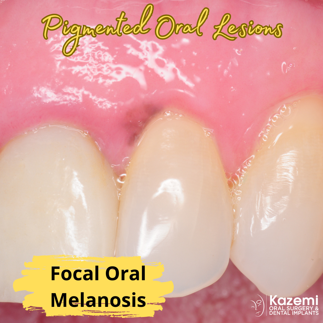
- Physiologic Pigmentation: Physiologic pigmentation refers to normal variations in pigmentation within the oral mucosa, which may be more prominent in certain individuals or ethnic groups. These pigmented areas are usually benign and do not require treatment.
- Drug-Induced Pigmentation: Some medications or substances can cause pigmentation changes in the oral mucosa as a side effect. For example, certain antimalarial drugs, chemotherapy agents, or heavy metals may lead to oral pigmentation.
It’s important for any pigmented oral lesion to be evaluated by an oral surgeon, for proper diagnosis and management. While many pigmented oral lesions are benign, some may require further investigation or treatment, especially if they show concerning features or are associated with symptoms. An excisional or incisional biopsy is indicated for definitive diagnosis.

Dr. H. Ryan Kazemi is a board-certified oral and maxillofacial surgeon in Bethesda, MD