In the realm of dentistry, accurate diagnosis forms the cornerstone of effective treatment planning and intervention. When it comes to identifying dental infections and bone loss, traditional peri-apical dental X-rays have long been relied upon. However, the advent of Cone Beam Computed Tomography (CBCT) has revolutionized diagnostic capabilities in dentistry, offering a myriad of benefits over conventional X-rays. This article explores the advantages of CBCT in diagnosing dental infections and bone loss, highlighting its superiority over peri-apical dental X-rays.
- Enhanced Visualization: CBCT provides three-dimensional images of the dental anatomy, offering unparalleled visualization compared to two-dimensional peri-apical X-rays. This comprehensive view enables dentists to accurately assess the extent of dental infection and bone loss, identifying even subtle changes in bone density and morphology.
- Precise Localization: With CBCT, dentists can precisely localize the site of infection or bone loss, facilitating targeted treatment planning. Unlike peri-apical X-rays, which often require multiple exposures to capture different angles, CBCT captures a single scan encompassing the entire region of interest, reducing patient discomfort and radiation exposure.
- Detailed Assessment of Bone Quality: CBCT allows for a detailed assessment of bone quality and quantity, crucial for evaluating the feasibility of dental implant placement and assessing the prognosis of compromised teeth. By visualizing bone density and morphology in three dimensions, CBCT enables dentists to make informed decisions regarding surgical interventions and treatment outcomes.
- Improved Treatment Planning: The comprehensive imaging provided by CBCT aids in meticulous treatment planning, enabling dentists to formulate customized treatment strategies tailored to the individual patient’s needs. Whether it involves root canal therapy, periodontal treatment, or implant placement, CBCT facilitates precise localization of pathology and optimal treatment execution.
- Reduced Diagnostic Errors: Compared to peri-apical X-rays, which may suffer from anatomical superimpositions and distortion, CBCT minimizes diagnostic errors by providing distortion-free, high-resolution images. This accuracy enhances diagnostic confidence, reducing the likelihood of misinterpretation and ensuring optimal patient care.
- Patient Education and Engagement: CBCT images offer patients a clearer understanding of their dental condition, fostering active participation in treatment decisions. By visualizing the extent of dental infection and bone loss in three dimensions, patients gain insight into the urgency and significance of recommended treatments, leading to improved treatment acceptance and compliance.
Cone Beam CT (CBCT) emerges as a game-changer in the diagnosis of dental infections and bone loss, outshining traditional peri-apical dental X-rays in several aspects. Its ability to provide enhanced visualization, precise localization, detailed bone assessment, improved treatment planning, reduced diagnostic errors, and enhanced patient education underscores its indispensability in modern dentistry. By harnessing the power of CBCT, dentists can elevate the standard of care, ensuring optimal outcomes and patient satisfaction in the management of dental infections and bone loss.
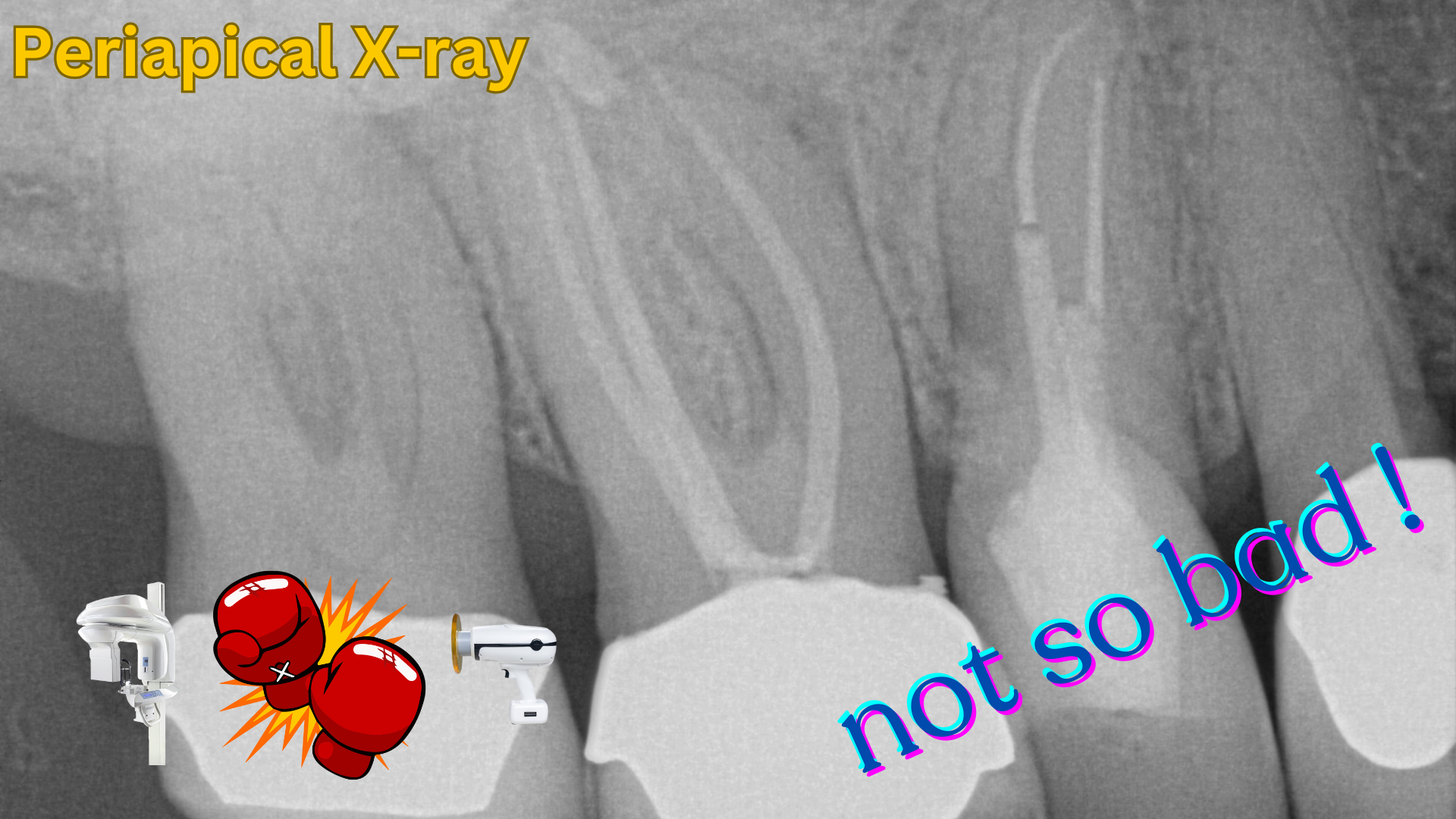 A peri-apical X-ray showing relatively normal level of bone and no notable pathology.
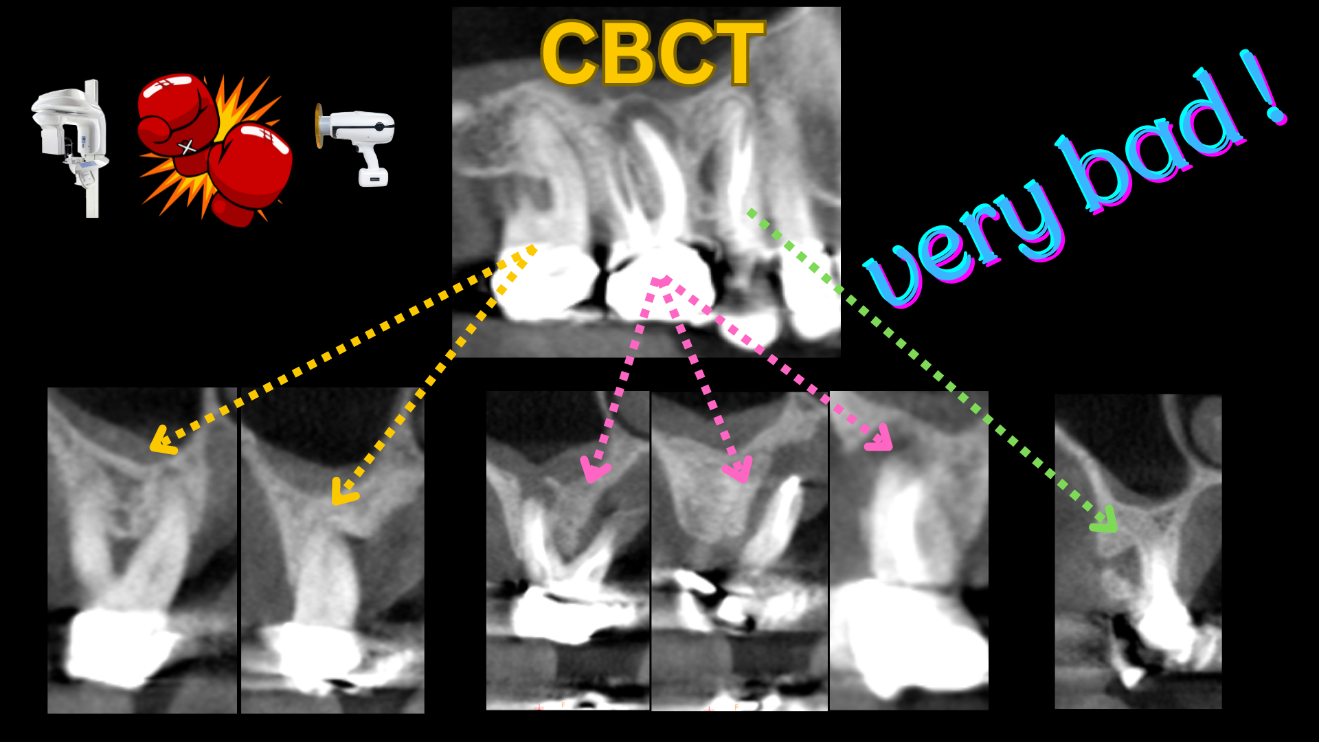
The same teeth imaged by a cone beam CT scan showing extensive infection, bone loss, perforation into sinus floor, and compromised foundation.
Dr. H. Ryan Kazemi is a board-certified oral and maxillofacial surgeon in Bethesda, MD