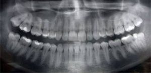
In order to achieve predictable results in wisdom teeth extraction, it is important that the oral surgeon utilizes appropriate diagnostic tools and methods specific to the planned procedure. For wisdom teeth extractions, this diagnostic tool is the panoramic X-ray (Panorex). Regular dental X-rays (periapical or bitewing) are inadequate for wisdom teeth because they do not provide the surgeon with a complete picture. Standard dental X-rays have a limited field of view and, therefore, rarely show the wisdom teeth in their entirety. A panoramic X-ray, on the other hand, provides a complete view of the jaw and teeth. The Panorex shows the teeth and roots and their stages of development. It also shows their relationship to adjacent teeth, sinuses, and nerve canals. The Panorex enables surgeons to accurately diagnose, plan, and safely treat wisdom teeth extractions.
In patients with deeply impacted wisdom teeth, or when there is evidence of oral pathology, such as jaw cysts, a dental CT scan may be recommended in addition to the Panorex. This helps the oral surgeon to further assess the position of the teeth, the size of related defects, and the proximity of vital structures, such as sinuses or nerves.
Knowing that teeth may change position or undergo pathological changes, it is imperative that the Panorex is taken within six months prior to the procedure. This ensures a quality diagnostic work up and predictable surgery results. Operations based on a non-diagnostic X-ray can lead to many complications and poor results.
When planning your wisdom teeth extractions, make sure to request a skilled oral surgeon and get proper X-rays. To learn more about wisdom teeth extractions, download Dr. Kazemi’s ebook: “The Wise Guide to Wisdom Teeth Extraction”

