 Dental impressions are an inherent part of dental implant replacement of missing teeth. They are required to capture the teeth anatomy for both diagnostics, as well as fabrication of restorations. Conventional impression techniques use trays and materials such as alginate or polymer compounds that when mixed transform from a flowable form to a more rigid state. The impressions are then poured in stone to create working models. Some may refer to this as the ‘stone-age’ technique!
Welcome to digital dentistry! Advanced technologies of cone beam CT scan and intra-oral scanners now enable us to provide total digital solutions for replacement of missing teeth and other restorative procedures. The benefits of digital implant dentistry include remarkable accuracy and precision, less number of patient visits, and shorter overall treatment time. Let’s take a look at how we use such technology for dental implant replacement of missing teeth:
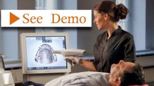
- Study models: Intra-oral scanners can be used to obtain digital impressions of the patient’s teeth and print study models for diagnostic purposes. The models help the surgeon and the restorative dentist understand patient’s bite scheme (occlusion) and design a restorative-based treatment plan.
- Dental implant planning: In preparation for dental implant placement, a cone beam CT scan is obtained and then merged with the intra-oral scan to create a complete 3-dimensional representation of patient’s teeth, gum tissue, and bone. Next, digital restorations are placed in this computer model to simulate the planned bite or occlusion. Then virtual dental implants are placed, each corresponding to the conceived restoration and the underlying bone. The surgeon can now easily select the optimal dental implant diameter and length and plan its proper orientation.
- Fabrication of a surgical guide: The information obtained from virtual implants and restorations is then used to fabricate a CAD / CAM surgical guide which helps the surgeon place the implants precisely according to the computer simulation.
- Fabrication of a custom abutment: Following healing of dental implants, special scan posts are attached to the implant, and a digital impression is obtained using the intra-oral scanner. This information is then shared with a laboratory where temporary or final abutments are fabricated with exact specs from the restorative dentist.
- Fabrication of temporary and final restorations: The final restorations are then digitally designed, milled, and returned to the restorative dentist for insertion.
Patients find this approach much more comfortable than traditional impression techniques, particularly those who have gag reflex and do not tolerate impression materials well.