 Most people don’t think about their sinuses unless they are dealing with a sinus-related illness, infection, or allergies. But did you know that the fact that your sinuses are located right above your upper teeth poses a potential problem like sinus perforation when having one of your upper teeth extracted?
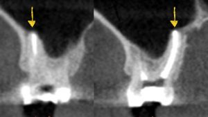
The anatomy of the sinus floor and its relationship to the upper teeth varies from person to person. In some people, the sinus floor is well above the roots of their teeth with bone separating the roots from the sinuses. While in other people the floor follows the roots more closely. In essence, wrapping around the roots with minimal bone between the sinus and tip of the roots (as shown in the photo). For those whose sinuses are very close to or even touching their tooth roots, there is a risk that the sinuses will be perforated when their tooth is extracted. This risk is greater if the tooth being extracted is infected or has an abscess at the tip of the root(s).
When considering an extraction of an upper tooth, if the conventional x-rays show that the tooth’s roots are near the sinus floor above, or if there are signs of an infection or abscess, then it is critical to obtain a pre-surgical cone beam CT scan (CBCT). A CBCT can assess the proximity of the roots to the sinus or assess the degree of existing defects that may lead to a sinus perforation following an extraction.
Sinus perforations, if not diagnosed and left untreated, can persist, leading to an oral-antral fistula—a continuous opening between the sinus and the mouth. Oral-antral fistulas can result in sinus infections as well as fluid drainage from the mouth to the nose. However, with a proper diagnostic CBCT, the surgeon and patient can discuss the likelihood of sinus perforation in advance and plan for its management before surgery. It is important to manage sinus perforation at the time it occurs to prevent it from progressing to a chronic oral-antral fistula.
A sinus perforation following a tooth extraction is managed using a three-layer approach. The first layer involves careful placement of collagen plugs or resorbable membranes at the junction of the root and the sinus opening. It is important to place this material carefully, so it does not get displaced into the sinus. In the next layer, bone graft particulate is packed gently into the sides, and not directly down, to avoid displacement into the sinus. In the third layer, another collagen plug or resorbable membrane is placed over the bone graft to keep it intact and protected in the socket. Finally, cross sutures are placed to stabilize the membrane over the graft and maintain its position.
Patients with sinus perforation are provided with specific instructions to follow for 2-3 weeks following the surgery to prevent any complications from developing. The gingival tissue, or gums, will heal over the bone graft in about 4-6 weeks, and the bone graft will heal completely in 4-6 months.