 One of the potential complications with wisdom teeth extraction is injury to the inferior alveolar nerve which can result in prolonged or permanent numbness of the lip and chin area or other sensory disturbances. The literature cites the incidence of wisdom teeth extraction-related nerve disturbance between 0.35 and 8.4%. While this complication is relatively rare, it is certainly a serious concern for patients and surgeons alike and its risks should be discussed in detail during consultation.
Nerve injury can occur during extraction of a lower wisdom tooth if it is in direct contact with the mandibular canal which contains the inferior alveolar nerve, a sensory third division of the trigeminal nerve. The close proximity of the lower wisdom teeth and the canal is a normal anatomical finding. However, in some patients the roots may be in direct contact with the nerve or have altered root anatomy (i.e. curves, hooks, dilaceration, etc) increasing the risk of nerve damage during extraction.
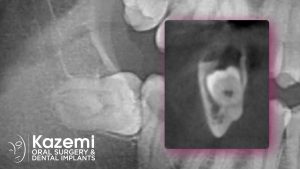
Panoramic X-rays are commonly used to assess the condition and position of the wisdom teeth before extraction. However, panoramic X-ray is a 2-dimensional image and does not provide precise information on exact relationship of the roots to the nerve canal. Because of this limitation, sole use of panoramic X-rays can be misleading in diagnosis and recommendations for wisdom teeth extractions. In circumstances where the roots ‘appear’ close to the canal, a cone beam CT scan should be obtained for proper visualization of the roots and the canal and definitive diagnosis and planning. In many patients, the panorex may show ‘roots on top or crossing the nerve’ but are actually separate when viewed on a 3-D cone beam CT scan.
There are two ways to minimize or prevent nerve injury and related lip / chin numbness during wisdom teeth extraction. The first is skilled use of minimally-invasive and atraumatic surgical techniques. This means the oral surgeon extracts the wisdom teeth with minimal manipulation- no excessive tilting, pressing, or twisting motions- but rather a gentle elevation of the tooth away from the canal. This way, even if the nerve and the roots are in intimate contact, the nerve remains intact during extraction. This is what I call the global precaution in wisdom teeth extractions: Gentle and atraumatic techniques in every patient with the assumption that the teeth and the nerve are in close proximity. The technique is discussed in the ebook, “The Wise Guide for Wisdom Teeth Extraction”.
The second is a modified technique known as partial extraction therapy. It is also referred to as coronectomy when performed for wisdom teeth. With this approach, about half of the tooth is removed with the roots intentionally left in place hence avoiding any chance of nerve trauma. The site heals normally and the root simply becomes calcified in its position. This procedure has proven to be a safe and predictable alternative with little post-operative complications when performed correctly. This technique should be used only when the roots of wisdom teeth and the mandibular canal have been definitively diagnosed to be in intimate contact or if the roots are located in close proximity with anatomical anomalies such as curves or hooks that may increase risk of nerve injury during extraction. It is contraindicated if the wisdom tooth has peri-apical (tip area of the roots) infection or mobility.
Partial extraction therapy is an effective technique for management of impacted or non-impacted wisdom teeth that present with close proximity to the inferior alveolar nerve. Speak to your oral surgeon for specific information on its use and indications in your case.
Dr. H. Ryan Kazemi is a board-certified oral and maxillofacial surgeon in Bethesda, MD