Cysts in the jaw bone are commonly related to impacted or malpositioned teeth. Such odontogenic cysts are often benign but cause extensive resorption of the jaw bone. One of the most common types of odontogenic cysts is dentigerous cyst which makes up about 20-30% of all jaw cysts. They often occur in the twenties to thirties and are considered quite rare in children.
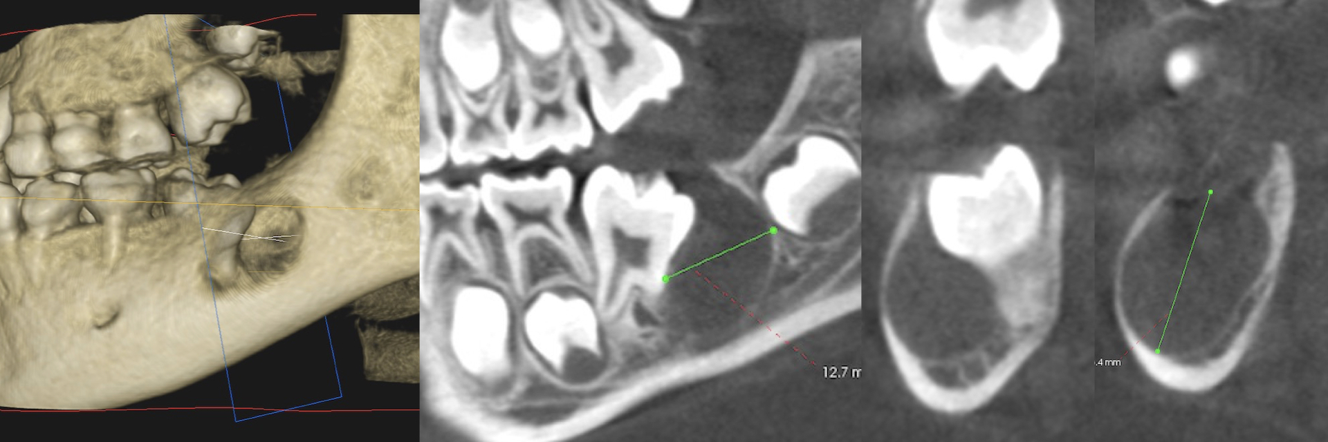
A 9-year old patient had presented with a delayed eruption of the lower first molar. X-ray showed an impacted molar with a radiolucent cystic lesion. The Cone Beam CT Scan (CBCT) demonstrated an extensive lesion extending to the full width and height of the lower jaw bone.
A histological diagnosis is critical for the definitive management of such large lesions. The partial biopsy with a small sample of the cystic lining confirmed a diagnosis of a dentigerous cyst.
The ideal treatment for dentigerous cysts consists of complete removal of the cyst (enucleation), extraction of the related impacted tooth, and bone graft to restore the bony defect. However, due to the patient’s age and the role of the first molar in supporting the bite, preservation of the related impacted tooth and close monitoring was suggested as an alternative approach.
The cyst is removed entirely, grafted, and closely monitored for two years. The protocol is clinical evaluation and CBCT at six months, one year, and two years. In the absence of recurrence, the tooth’s eruption is monitored during growth and development, considering exposure and orthodontic-assisted eruption as necessary. If the cyst recurs, the tooth and the recurrent cyst will be likely removed and grafted.
Dr. H. Ryan Kazemi is a board-certified oral and maxillofacial surgeon in Bethesda, MD