Exposure and orthodontic eruption of impacted canines is an effective and predictable treatment when performed at early ages, typically between ages of 12-14. However, in older patients such procedures are significantly less predictable. The bone becomes increasingly calcified and more dense and hence the impacted canine becomes more anchored in place and less responsive to orthodontic eruption forces. Another common reason for poor response in older patients is ankylosis with or without external root resorption.
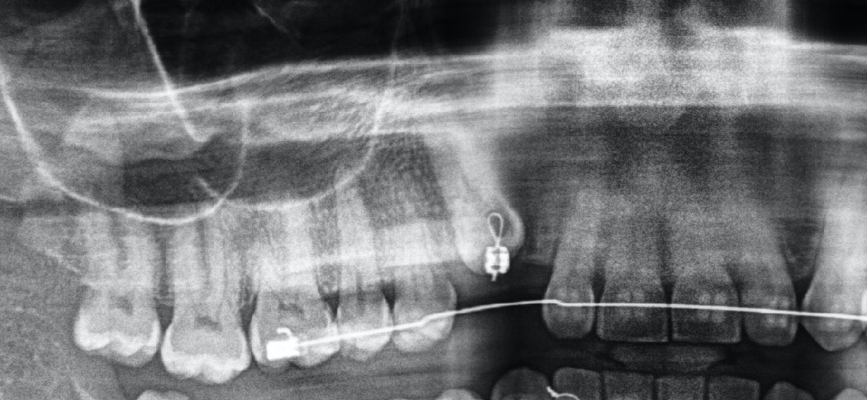
This 55 year old patient was in active orthodontic treatment for over 2 years with little movement of the impacted canine. The initial diagnostics and monitoring was done using only 2-dimensional panoramic x-rays as seen below. The panorex shows the impacted canine but does not provide any other helpful information regarding its condition and surrounding structures.
With lack of tooth movement, patient sought a second opinion with us to determine its cause and possible treatment options to help further its eruption. A cone beam CT scan (CBCT) was recommended to assess the anatomy of the canine, its trajectory, and rule out possible residual roots or other structures that may have impeded its eruption. A CBCT is a remarkably valuable diagnostic imaging tool that should be done prior to initiating canine exposure and eruption as well as when the eruption process is either halted or very slow.
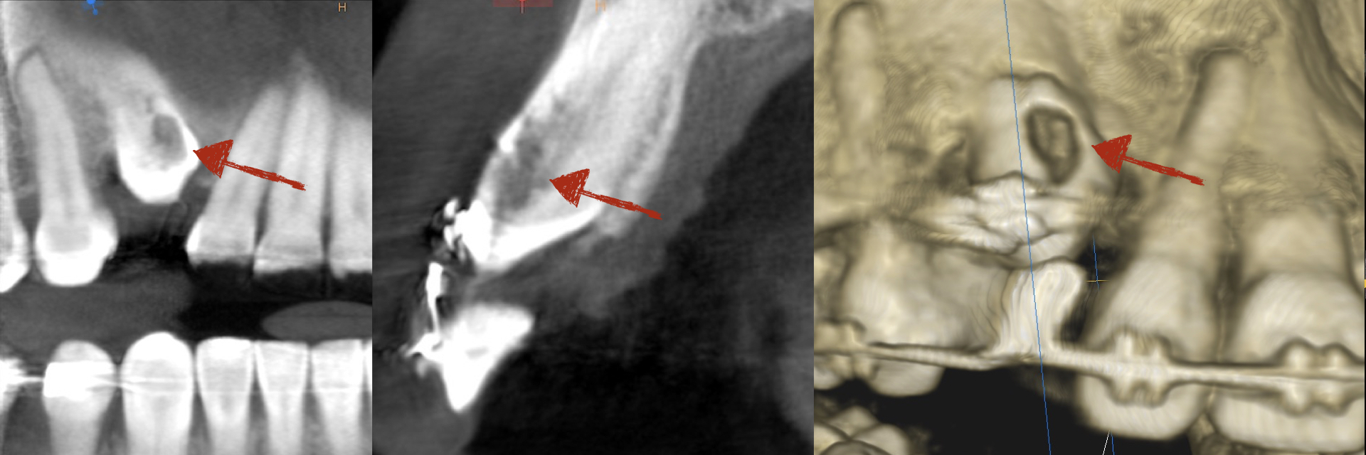
The CBCT demonstrated an extensive external resorption involving 60% of the tooth’s width at the crown level. External resorption is commonly associated with ankylosis, a common cause of non-responsive impacted canines to orthodontic eruption in older patients. Even if the tooth was successfully erupted, it is non-restorable and guarded and would require extraction regardless. The treatment plan for managing this patient includes:
- Establish the proper space with orthodontics and retain the position of adjacent teeth.
- Extraction and site bone graft to restore and preserve bone and soft tissue
- Delayed implant placement and restoration
This case demonstrates the importance of 3-d imaging in diagnosis and team planning to make sure treatments are appropriate and effective. A close communication between the orthodontist, restorative dentist, and surgeon is vital in planning and execution of such treatments to assure optimal success and outcome for patients.
Dr. H. Ryan Kazemi is a board certified oral and maxillofacial surgeon in Bethesda, MD