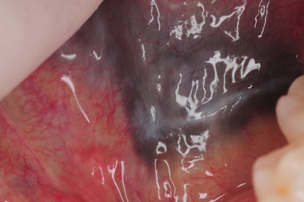
Amalgam tattoo is a benign lesion which appears as black or blue lesions in the mouth. It is caused by embedding of fine particles during removal of the amalgam restorations which can be prevented by use of a rubber dam. They are typically non-tender and do not change their characteristics or size over time.
Since such pigmented lesions in the mouth may have similar appearance to oral melanotic macule or possibly melanoma, an incisional or excisional biopsy is recommended for definitive diagnosis. This involves a minor surgical procedure where the tissue sample is obtained and sent to an oral pathologist for histological diagnosis. Amalgam tattoo can be removed completely if it is in an aesthetic visible area. The recovery from this relatively minor biopsy procedure is often quick with little discomfort.