 [By Lifetime Dental Implants at Kazemi Oral Surgery in Bethesda, MD / Washington DC / Virginia]
Diagnostic imaging is an important part of implant dentistry and is used to ensure safe and predictable treatments. Cone Beam CT Scan (CBCT) is an in-office imaging technique that provides 3-dimensional images of the jawbone, teeth, and surrounding vital structures that are important in planning the placement of dental implants. With its low cost, relatively low radiation, and ease of use, it is now a routine imaging technique for all dental implant treatments. Placement of dental implants using conventional dental X-rays is no longer an accepted practice and is highly risky.
There are five major benefits of cone beam CT scan (CBCT) for dental implant planning and placement:
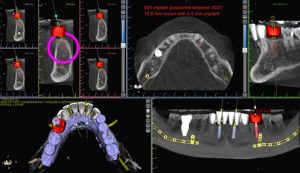
- Precision placement of implants in the bone: CBCT allows the surgeon to accurately measure and localize the available bone. Using 3-D software, we do a virtual implant placement with precise position and sufficient bone coverage. If bone is deficient, it is augmented using various bone grafting techniques. The key is to place the implant where it best supports the restoration while keeping it in healthy and abundant bone.

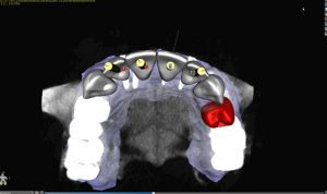
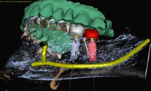
- Proper orientation of the implant with its overlying restoration: A CBCT is merged with an optical scan of the patient’s teeth (digital impression) to create a complete bone, teeth, and soft tissue virtual model. The dental implant surgeon and the restorative dentist then, in collaboration, design the perfect bite and precise position of the implants to support the planned restorations. This prevents misaligned implants, which may be difficult or impossible to restore, and avoids poor aesthetics and function.

- Prevention of injury to nerves: Using the CBCT, the surgeon maps out the path of the sensory nerves in the jawbone and selects the right implant length. Conventional X-rays are flat and distorted and are poor diagnostic images for predicting the position of the nerves. Nerve damage from dental implant placement results in partial or complete numbness of the lip and chin area, which can be potentially permanent. CBCT is a mandatory imaging technique to prevent this serious complication.

- Prevent implant penetration into the sinus: CBCT provides an accurate picture of the maxillary sinus and its position in relation to the available bone. The surgeon can make an accurate measurement and select the right implant length to avoid puncturing the maxillary sinus. Penetration of the maxillary sinus can lead to sinusitis or other inflammatory conditions. The surgeon can also plan for necessary bone grafting if there is insufficient bone to support the implant. Conventional X-rays are highly inaccurate for these purposes and do not provide the information necessary for the safe placement of dental implants in the back of the upper jaw and in the proximity of the maxillary sinus.

- Selection of the right size implant for optimal support: The longevity and success of dental implants require maximal integration and stability in the bone. CBCT allows the surgeon to measure the available bone and select the widest and tallest implant appropriate for the site. This, in turn, helps to support the high bite (occlusal) forces and avoid potential failure from overload. Implant size selection should not be guesswork! Implant selection is made based on precise measurements, biological requirements, bite scheme, and individual patient needs.
With the clear benefits it provides by offering safer and more predictable outcomes, CBCT should be a mandatory diagnostic imaging for every implant treatment. Not using CBCT for planning is unwise for the surgeon and creates unnecessary risk for the patient.
