Case report
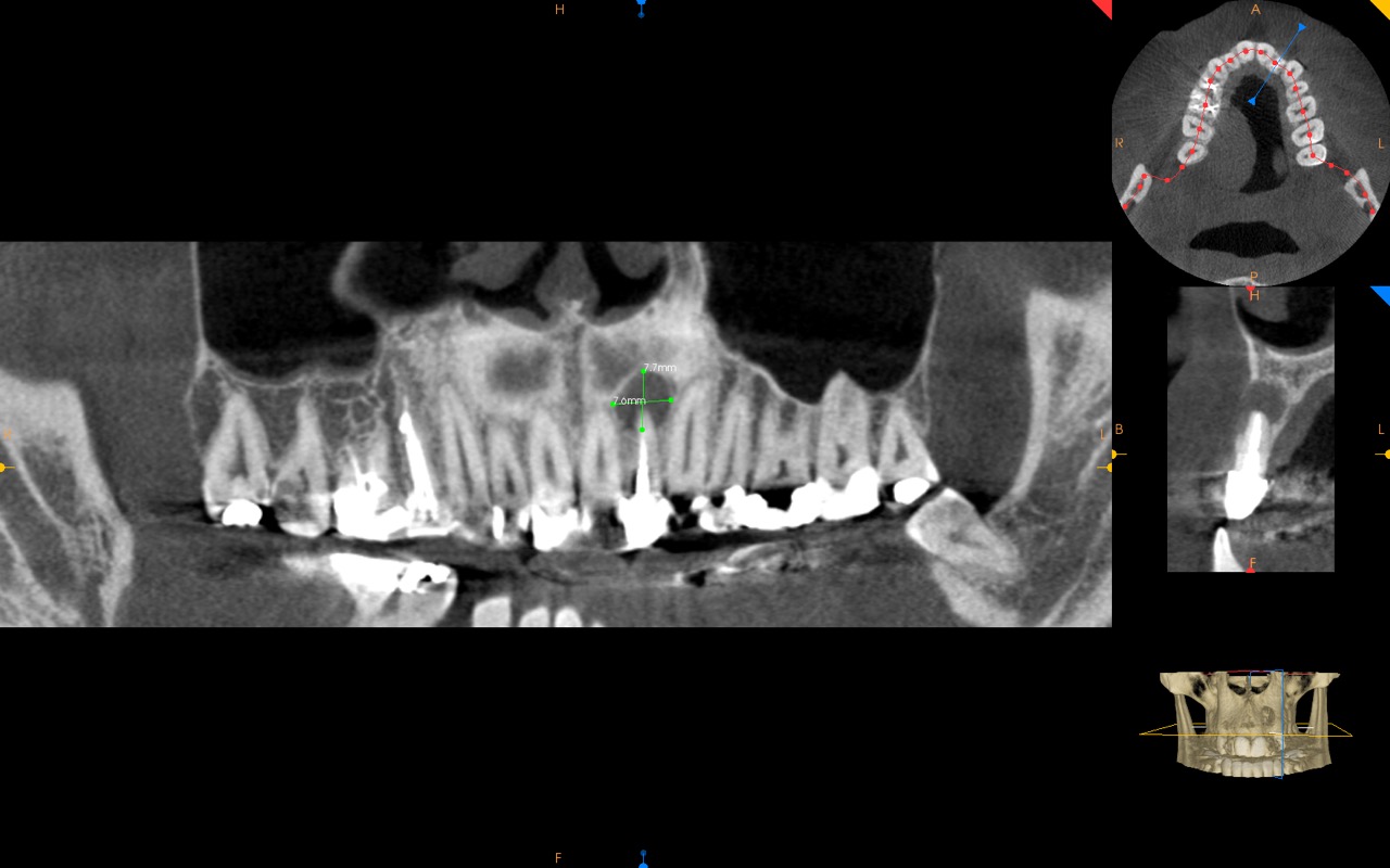
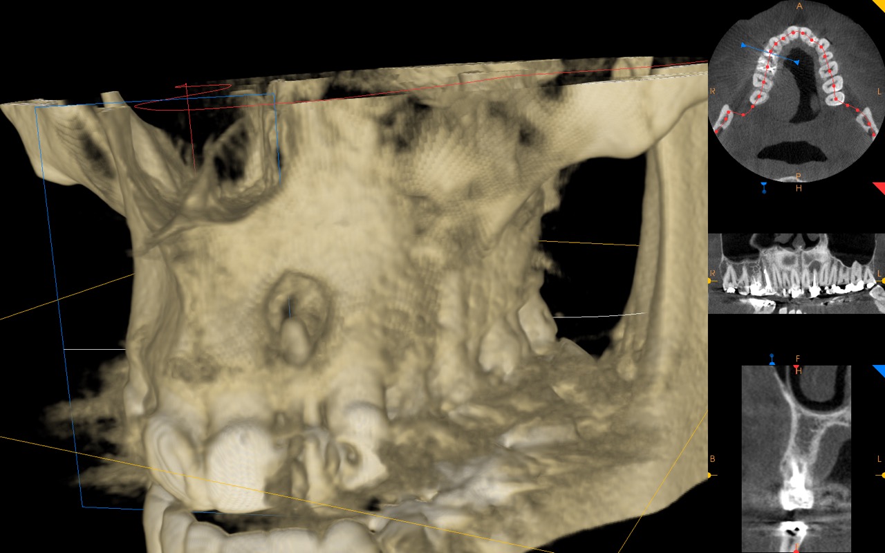
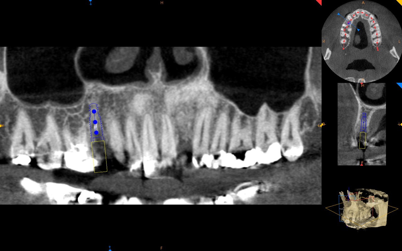
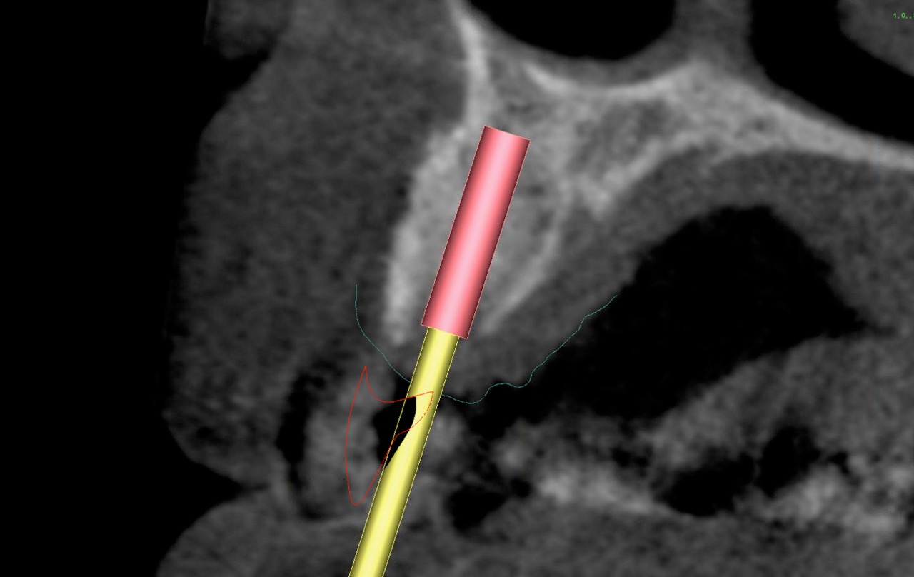
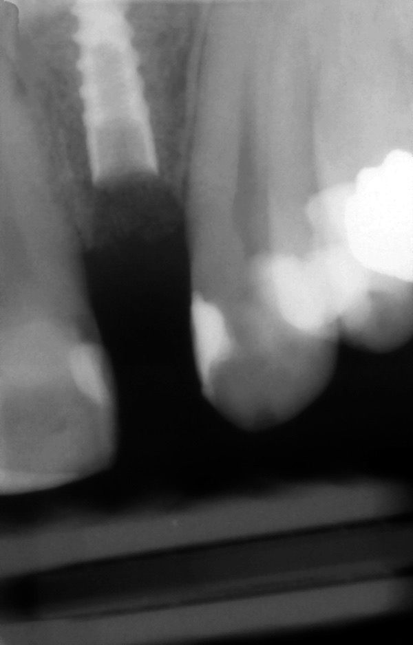
In this case report, we present a patient with a large periapical cyst involving tooth #10. A CBCT was obtained demonstrating the large cyst and bony defect with missing labial architecture of the bone. We describe site augmentation and then planning for dental implant placement using total digital workflow.

Treatment
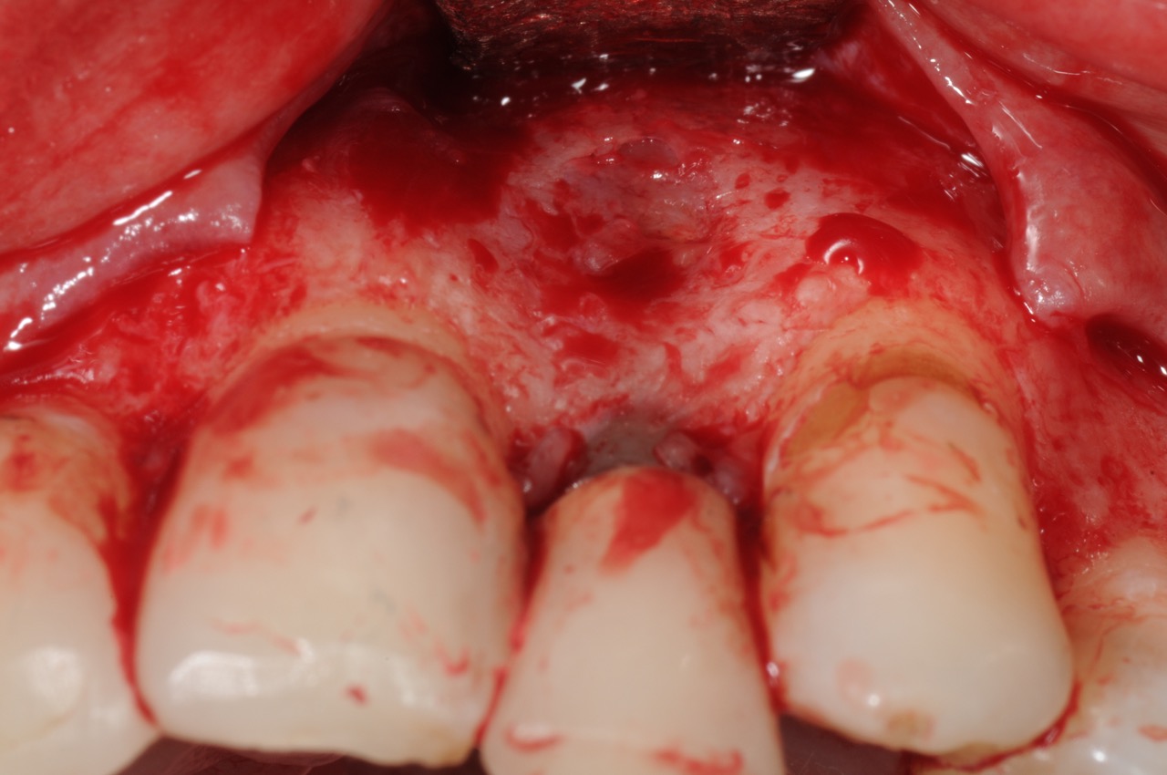
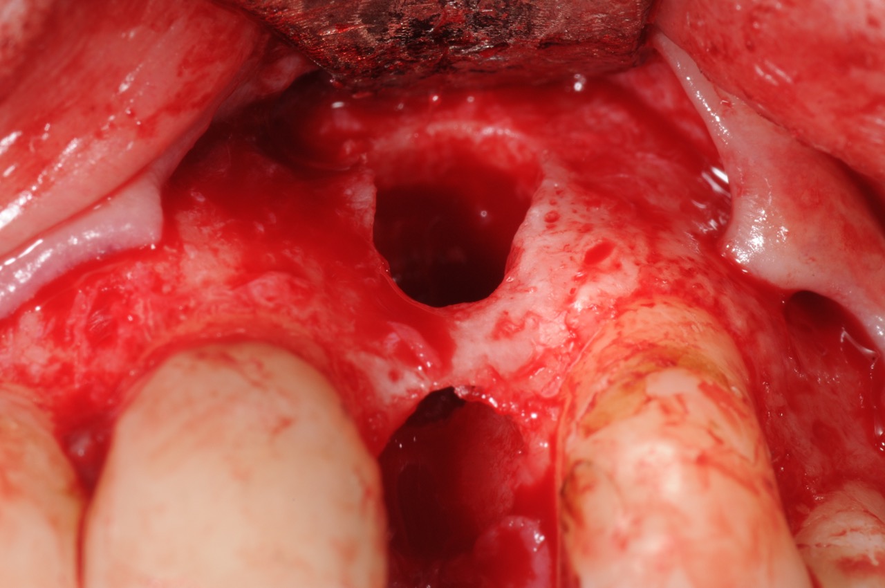
- The tooth was extracted and grafted to regenerate missing bone and then allowed 6 months to heal.
- A transitional removable prosthesis was provided as interim.
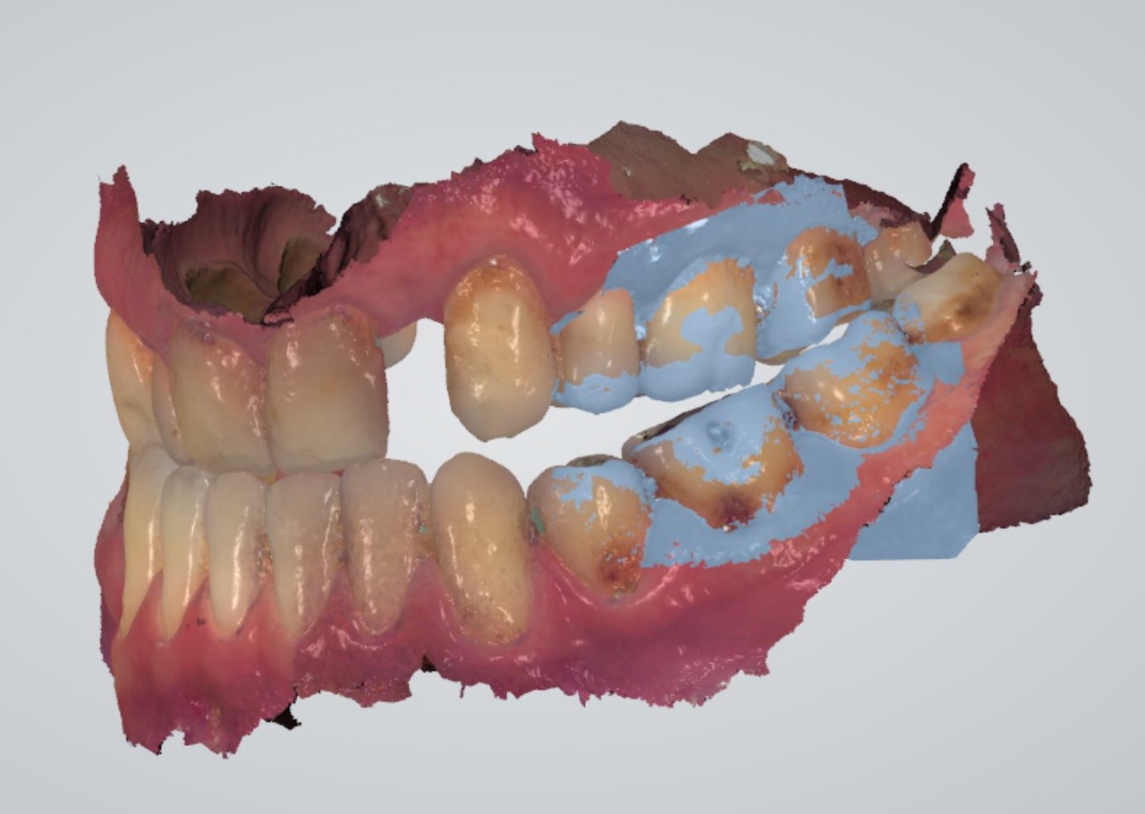
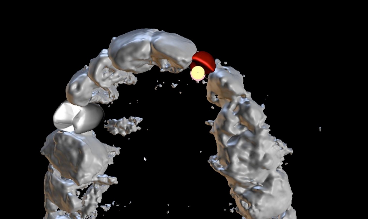
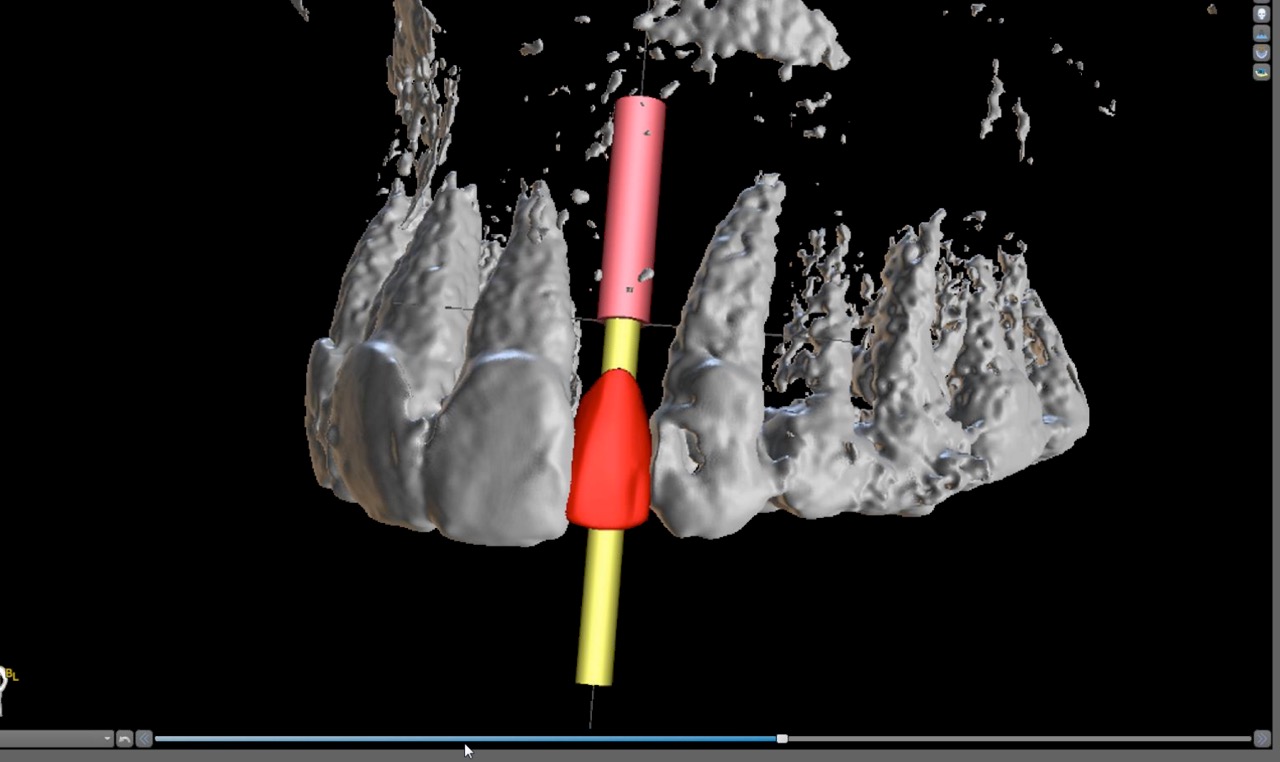
- For implant planning, a CBCT was obtained along with digital impression using a scanner. A 3-D computer model created and the virtual implant was placed. Due to the very narrow size of the bone, a 3.0 mm implant was planned and oriented precisely based on digital wax-up of the crown.
- The wax-up and plan was reviewed with the restorative dentist and approved.
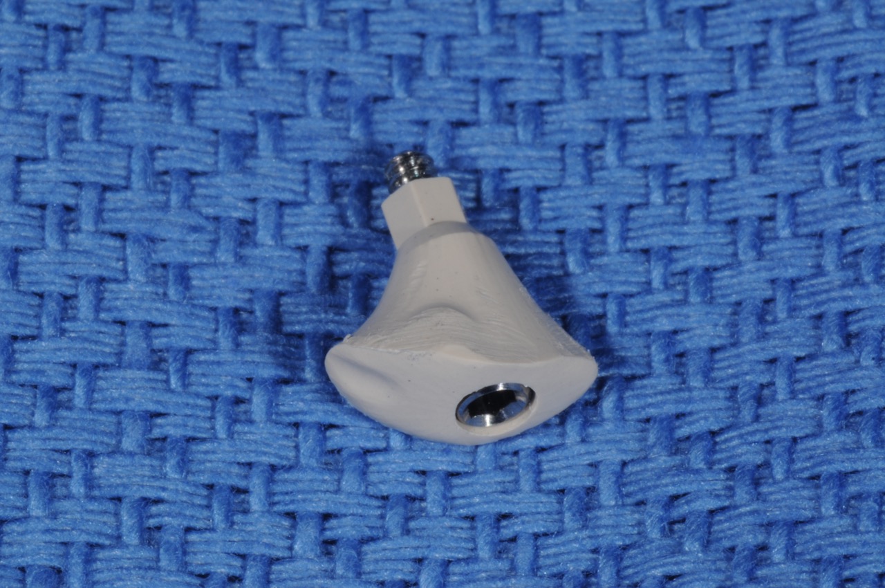
- A CAD/CAM surgical guide and a customized healing abutment were fabricated from the digital workup.
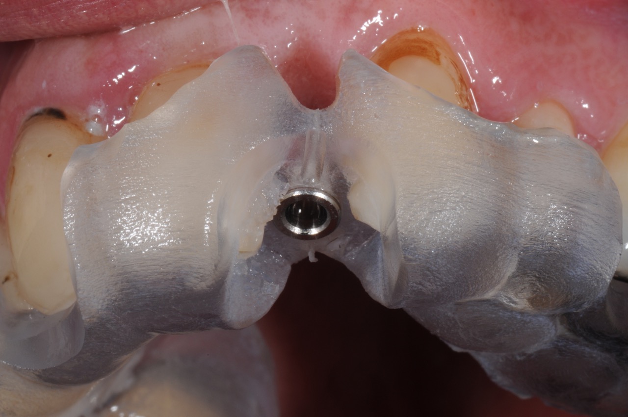
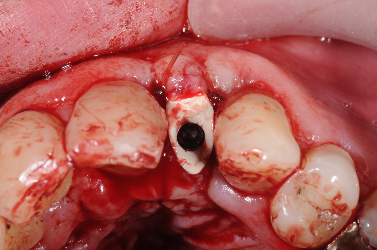
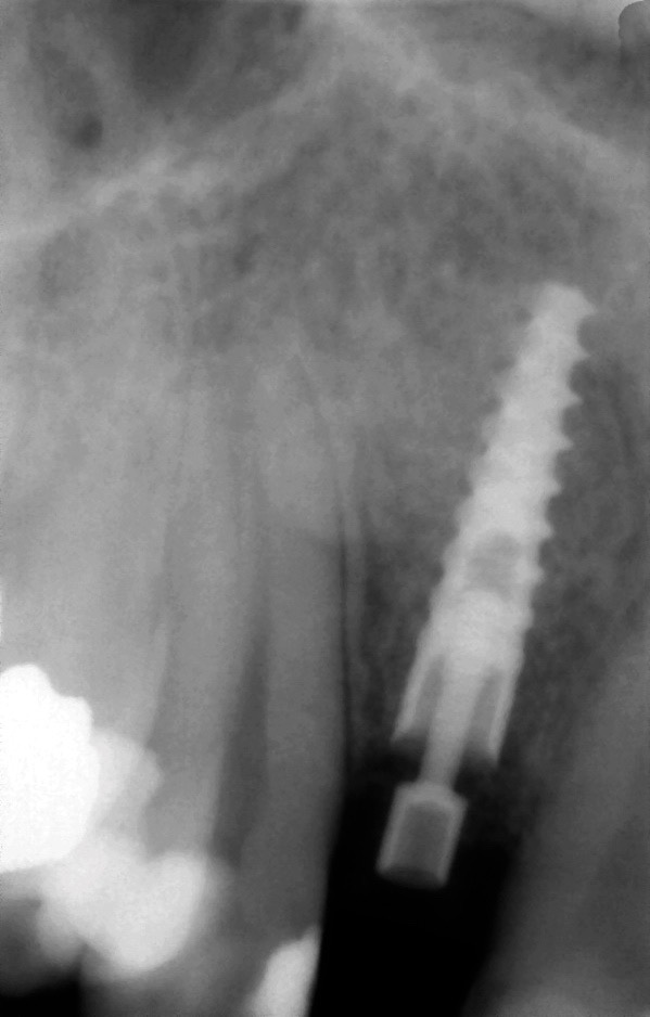
- A Ritter 3.0 narrow diameter implant was precisely placed using CAD/CAM surgical guide.
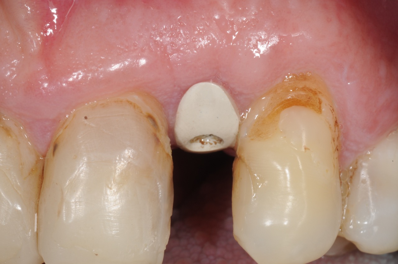
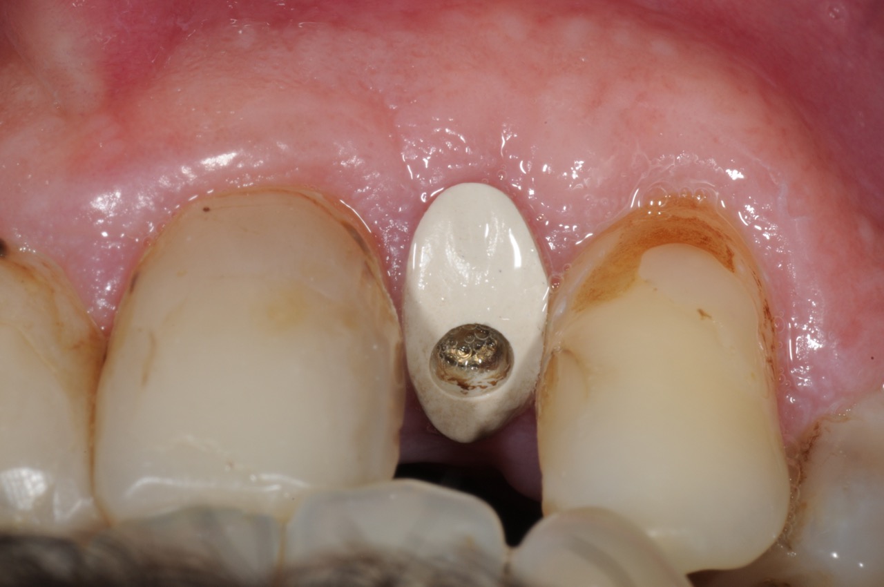
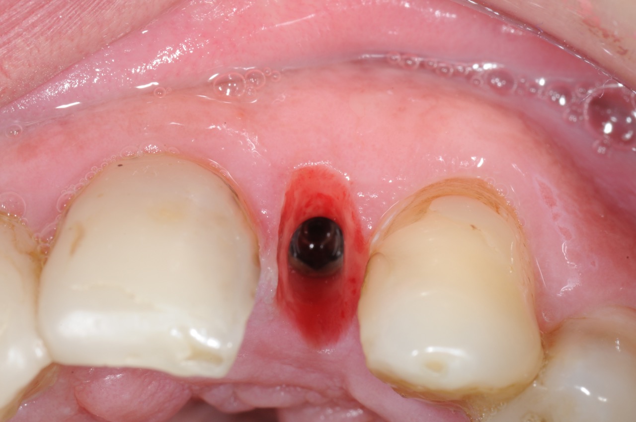
- The customized healing abutment was placed to design and form the proper gingival contour.
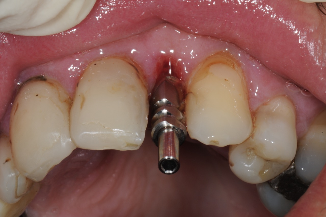
- Following 3.5 month healing, an impression of the implant was obtained using conventional impression post.
- A customized abutment and crown were fabricated and placed.
Success factors
- CBCT for proper diagnostics.
- Atraumatic tooth extraction.
- Proper site development with bone graft.
- 3-dimensional digital work up with precise implant position planning.
- CAD / CAM surgical guide for precise implant placement.
- Customized healing abutment to form proper gingival contours.
- Customized abutment and crown for optimal aesthetics.
- Close collaboration between the surgeon and the restorative dentist.
Photos
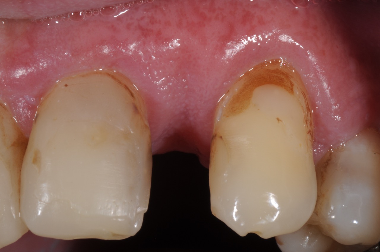
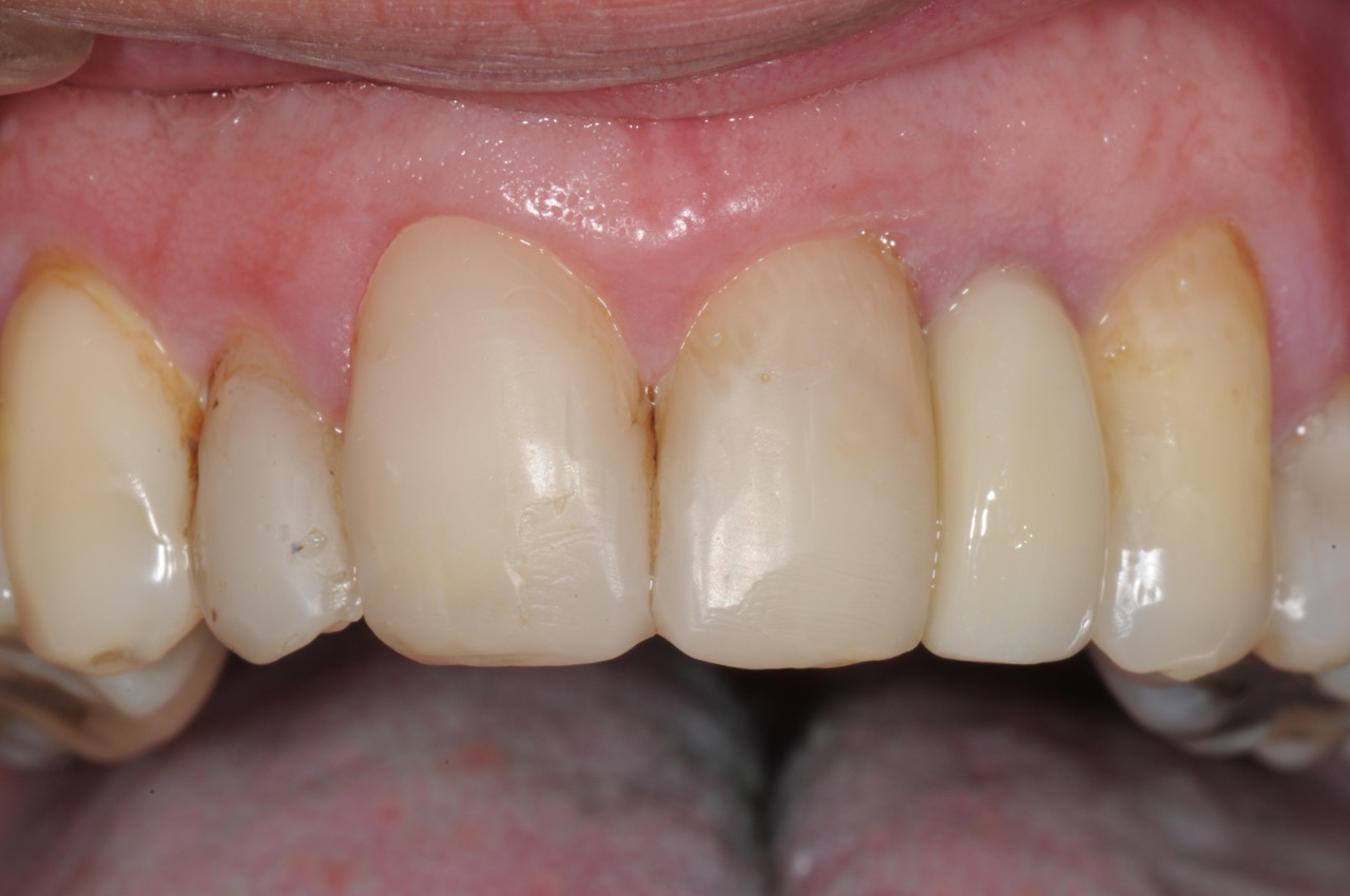
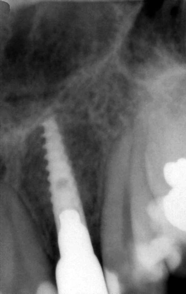
Before & after

