The Story:
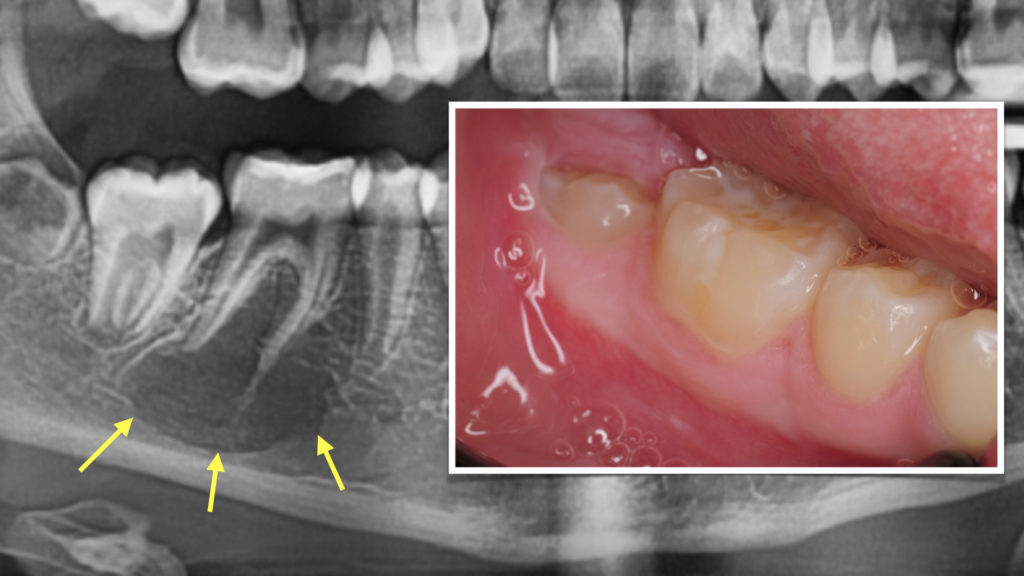
This 10 year old patient was noted with a dark area on her X-ray during routine dental examination. She had no symptoms or signs of any problems. The X-ray shows a well-defined radioleucent area in the right lower jaw surrounding the root of the lower first molar. Patient was referred to us for oral surgery and oral pathology evaluation.
Diagnostic Imaging:
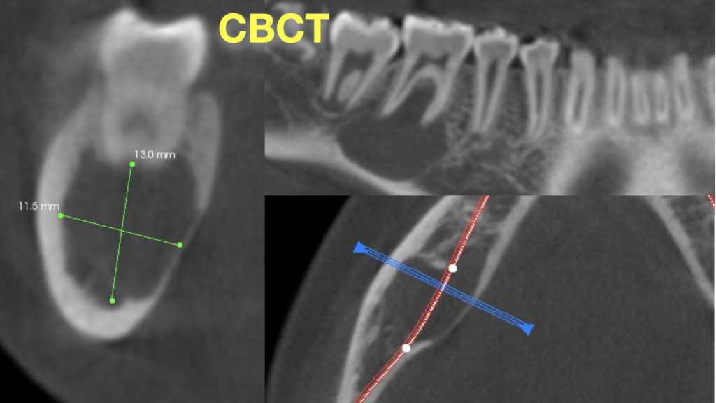
The first step was to obtain a cone beam CT scan (CBCT) to assess the size and presentation of the lesion. It was noted to be relatively large, but well-contained. Next, the molar was tested for vitality and noted to be normal. This is a critical test to make sure the root is healthy and the lesion is not an abscess that can have a very similar presentation. With these findings, the differential diagnosis included traumatic bone cyst.
Treatment:
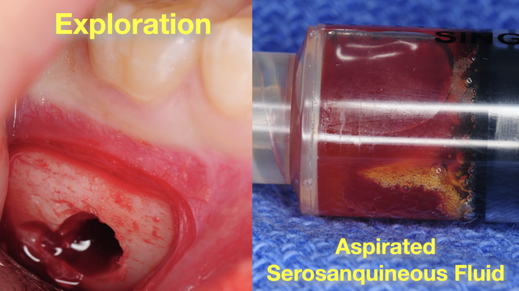
Through a small incision, the lesion was accessed and a small bony window was created. A serosanquineous (blood and blood serum) was aspirated. There was no lining within the cyst. These two findings confirmed the diagnosis of traumatic bone cyst. Incision is closed with resorbable suture.
Such lesions typically heal naturally following exploration procedure. An X-ray follow up in 6 months and 12 months is suggested to assure complete bony fill and healing.