The Story:
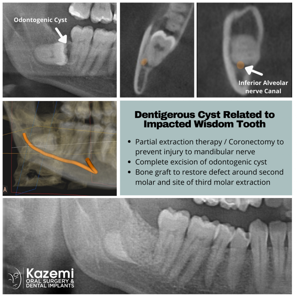
This patient was experiencing pain with her lower right wisdom tooth. Upon conventional X-ray, a cystic area was noted associated with the impacted wisdom tooth and the adjacent second molar. A 3-D cone beam CT scan (CBCT) was done for better imaging and assessment of the extent of the cyst. It was noted to be located between the wisdom tooth and the second molar. The 3-D dental scan also showed very close proximity of the wisdom tooth roots to the inferior alveolar nerve in the mandible (lower jaw bone). The treatment plan proposed included:
- Partial extraction therapy / coronectomy of the wisdom tooth to prevent injury to the nerve
- Complete removal of the cyst and clean the roots of adjacent second molar
- Bone graft along with platelet rich fibrin to restore the bony defect created by removal of the impacted wisdom tooth and the cyst and provide better support and improved tissues around the second molar.
- IV sedation for optimal comfort.
Treatment:
Under IV sedation, the impacted wisdom tooth was removed using partial extraction therapy (PET) also known as coronectomy. This allows intentional retention of the root tip area to avoid injury to the nerve. Next the cyst was completely enucleated and sent for histology / oral pathology study. The distal of the second molar was cleaned and scaled to prepare for bone graft. Bone graft material was combined with platelet rich fibrin obtained from patient’s own blood and then placed to restore the defect. It was covered with a resorbable membrane that provides for guided bone regeneration and followed by closure of the gum tissue.