The Story:
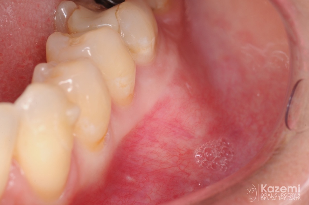
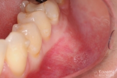
During routine dental exam and X-rays, the patient was noted with dark areas near roots of lower teeth. Patient was seen by an endodontist for evaluation and was noted to have vital teeth. She was then referred for evaluation of the jaw bone. Clinically, patients showed an area of expansion of the lower left area near the first molar region. Patient had no pain or any other symptoms.
Diagnostics:
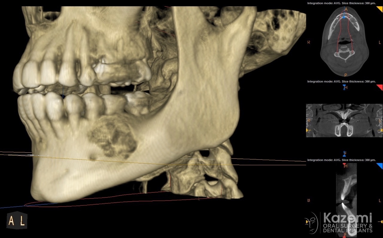
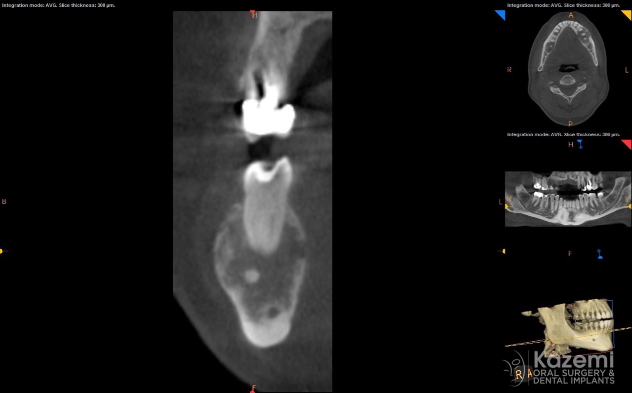
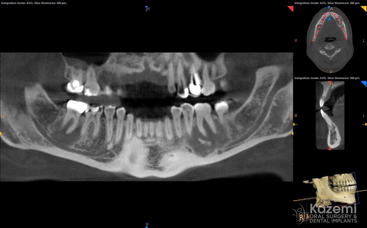
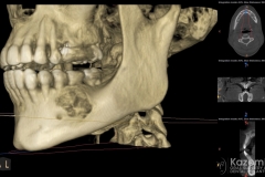
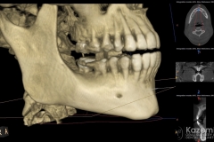
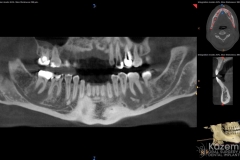
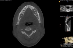
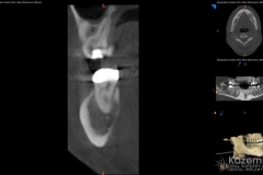
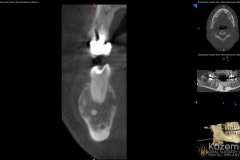
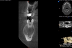
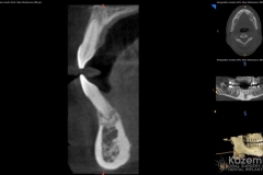
A complete cone beam CT scan (CBCT) was obtained showing areas of dark ‘cystic’ regions on both sides of the lower jaw as well as the front teeth. The teeth were all vital and with great periodontal attachment and stability.
Differential Diagnosis:
Based on the presentation of the bilateral bony lesions, vitality of the teeth, and absence of any symptoms, as well as the radiographic appearance, the condition is consistent with focal cemento-osseous dysplasia. This is a benign fibro-osseous condition. No treatment is required and only close follow-up is recommended as the condition can progress to the florid variation. Biopsy or other surgical interventions may result in osteomyelitis and hance should be done, if necessary, with caution and reviewing its risk and benefits. If patient becomes symptomatic or develops an infection, then surgical intervention such as a biopsy may be considered.
