The Story
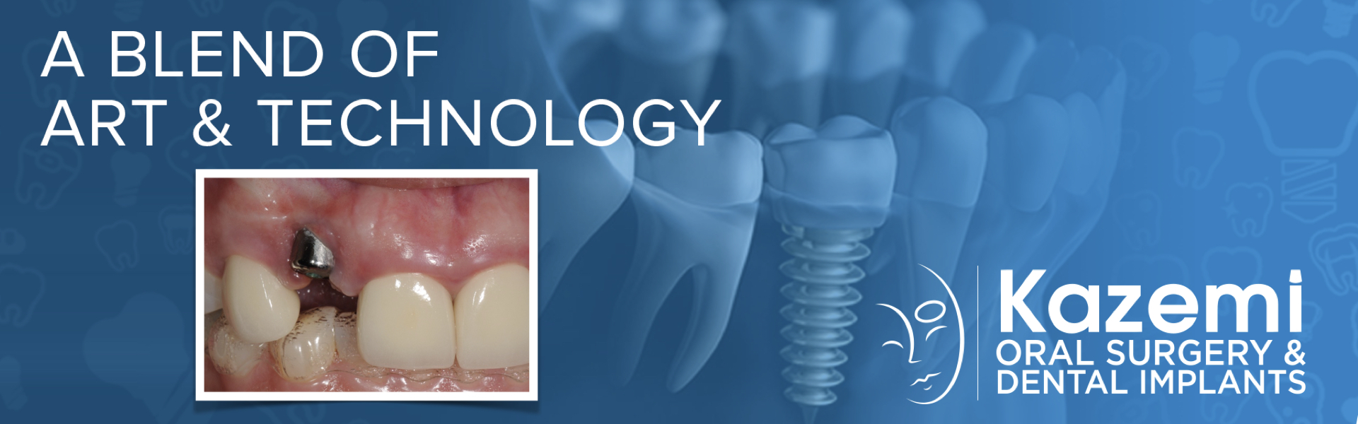
This patient presented to us for evaluation of her receding gum tissue following placement of a dental implant by her dentist. The dental implant was placed with no 3-d work-up or surgical guide. The examination and CBCT (cone beam CT scan) showed excessive angled implant position and no bone on its outer aspect. The gum tissue recession was due to lack of bone and also excessive tilting. Restoration of the implant would result is a significant aesthetic compromise. Gum or bone tissue grafting is ineffective in management of such a condition, therefore decision made to remove the implant , restore tissue, and then replace with new implant using digital / computer-assisted planning.

The Treatment
- Dental implant was removed using reverse torque explantation technique and platelet rich fibrin was placed for soft tissue healing and maturation
- The site was allowed to heal naturally bor 2.5 months while the gum tissue regenerated itself
- A CBCT (cone beam CT scan) was done to assess bone quantity. A bone graft was performed to restore the missing horizontal bone and allowed to heal for 6 months
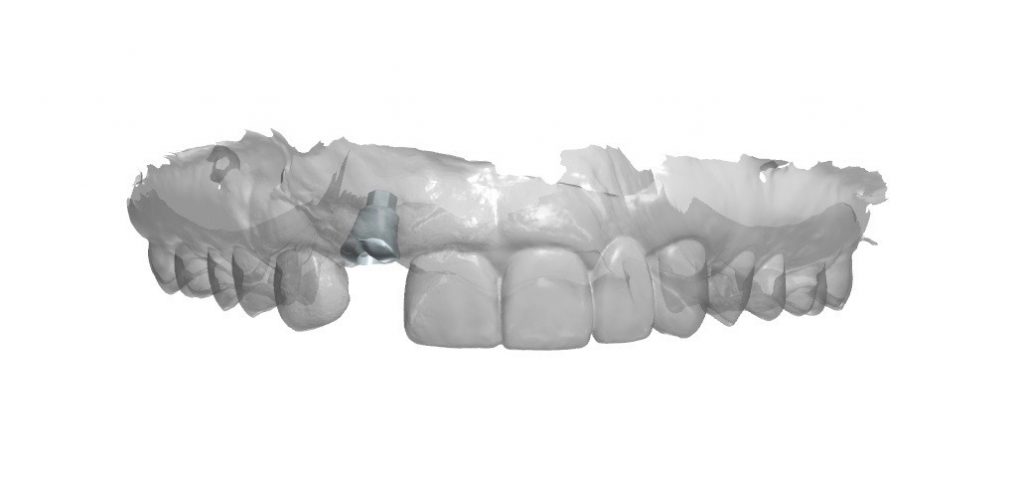
- For implant planning, a CBCT was obtained along with digital impression using a scanner. A 3-D computer model created and virtual implant was placed. Due to very narrow size of bone, a dental implant was planned and oriented precisely based on digital wax-up of the crown.
- The wax-up and plan was reviewed with the restorative dentist and approved.
- A 3-D printed surgical guide and a customized healing abutment were fabricated from the digital workup
- The customized healing abutment was placed to design and form the proper gingival contour.
- Following 3.5 month healing, an impression of the implant was obtained using conventional impression post.
- A provisional restoration supported by implant was designed and placed for 3 months to create the right soft tissue architecture
- A customized final abutment and crown was fabricated and placed.
Key Factors in Success
- CBCT for proper diagnostics
- Atraumatic implant removal
- Proper site development with bone graft
- 3-dimensional digital work up with precise implant position planning
- 3-d printed surgical guide for precise implant placement
- Customized healing abutment to form proper gingival contours
- Customized abutment and crown for optimal aesthetics
- Close collaboration between surgeon and restorative dentist.
Photos


Before & after