Dental implants have become the optimal treatment option for replacement of missing teeth. They have 98% + success rate and have allowed many patients to achieve normal eating and smiling without the irreversible damaging effect of other alternatives such as bridges or dentures.
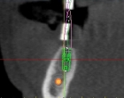
To achieve optimal functional and aesthetic results with dental implants, it is crucial that the patient has adequate bone and gum tissues and the implants are positioned properly in all dimensions. Utilizing readily available office CT scans, it is now possible to obtain cost-effective cross sectional images of the patient’s jaws that can then be converted to 3-dimensional images. These images accurately reveal the dimensions of the existing bone as well as the vital anatomical structures in proximity, such as nerves and sinuses. Often, the dentist first makes a special prosthesis (embedded with barium sulfate) that takes into account the final bite and teeth positions. The patient then wears this prosthesis during the CT scan creating radiological marks on those images. These marks represent the future sites of the crowns and the proposed positions of the dental implants, a critical tool in initial diagnosis and planning.
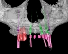
Utilizing special software, we then convert the obtained CT scan into 3-dimensional computer images that give us a 360 degree view of the patient’s jaw, available bone, its anatomy, and the surrounding vital structures. Next, we place virtual dental implants in their previously determined and planned positions guided by the marked sites on the CT. The result is a computer model demonstrating the existing bone anatomy, vital structures, and accurately positioned dental implants that will eventually support the patient’s teeth.
This 3-dimensional computer model is then used to fabricate a surgical guide or stent that translates the implant positions in the computer to a working guide that will be used by the oral surgeon during placement of the implants. These custom made guides are extremely well fitting, allowing the surgeon to easily and accurately place the implants in their appropriate positions. Once healed, the implants can easily be restored by the patient’s dentist knowing the foundations (i.e. dental implants) are in the right place and orientation.
We are utilizing this technology on many patients, allowing us better diagnosis, less complications, more accurate implant positioning, shorter and less invasive surgeries, and enhanced results that meets the patients’ goals.
The Steps:
- Your dentist takes models from your teeth. The laboratory will simulate the proposed final crown or bridge on the models. The laboratory then makes a splint that fits over your teeth. The teeth in the splint contain a special marker that allows it to show up in the CT scan
- Your dentist will then see you to check the splint for accuracy.
- An icat (CT scan) is done with the splint in your mouth.
- The CT data is then reformatted into 3-dimensional images.
- A special software is used to upload the images for computer simulation of the proposed implants. The marker shows up on these images, allowing the surgeon to accurately position the implants where they need to be in order to support the overlying crown or bridge.
- The final computer work up is then submitted to a laboratory to make surgical guides. These guides translate the positioning of the implant in the computer work up to a surgical splint to help the surgeon replicate it on the patient.

