Case report
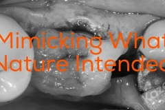
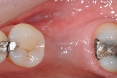
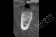
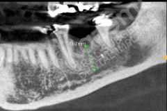
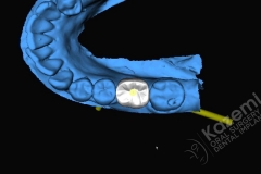
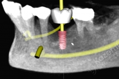
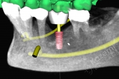
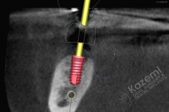
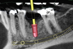
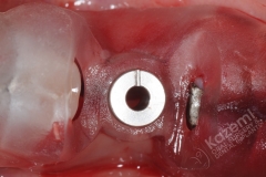
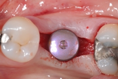
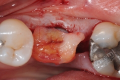
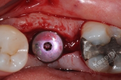
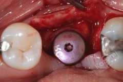
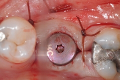
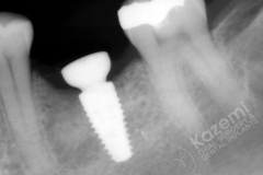
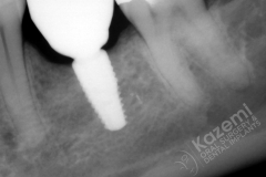
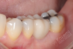
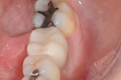
This patient had an extraction of lower molar with site bone grafting. So at time of implant placement had excellent bone foundation. However there was moderate deficiency of soft tissue (gingiva) making it look thinner at the top of the ridge. The treatment plan included 3-D computer assisted planning, implant placement with surgical guide, soft tissue (gingival) graft at time of implant placement, and restoration with custom abutment and crown. The outcome was a functional and aesthetic restoration with proper contours and tissue support.
Treatment
1. Diagnostics were obtained: CBCT and optical scan for 3-D planning
2. Surgical guide fabricated from 3-D work up using 3-D printing
3. Selected dental implant was placed using the guide.
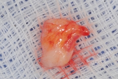
4. Soft tissue grafting was done from palate
5. Following 4 months of healing, a custom abutment and crown were fabricated and placed.
Success factors
1. The diagnostic work up began with the end in mind.
2. 3-D digital work flow for diagnostics and planning
3. A proper surgical guide allowed accurate placement of the implant.
4. Site development with soft tissue graftng. This allowed proper tissue topography and emergence.
