The Story:
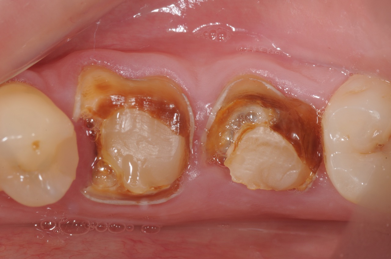
This success story demonstrates how we use the digital workflow for complete diagnostics and planning for precise dental implant placement. This patient presented with two non-restorable lower molars due to severe caries. After a thorough evaluation the diagnostic is to extract the teeth and plan for dental implant placement.
Treatment:
During the first procedure, Dr. Kazemi extracted the teeth using an atraumatic technique and grafted the site to augment and preserve the bone architecture. Following 5 months of healing, a cone beam CT scan and optical scan impressions are performed to prepare for 3-D computer-assisted planning.
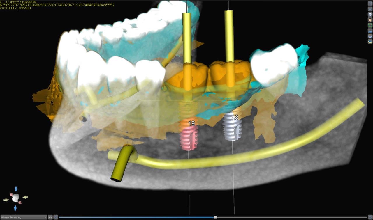
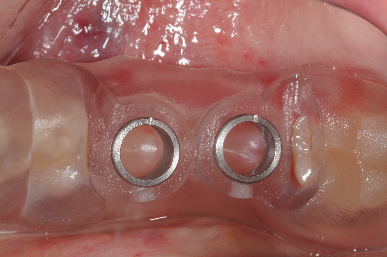
With precise 3-D planning, a CAD / CAM surgical guide is fabricated to allow the surgeon to place the implant in precise and planned position.
Final crowns are fabricated and supported by the implants.
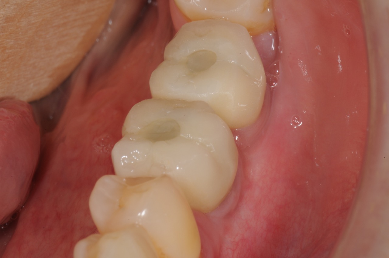
Photo Gallery (Click on first photo to start):


Keys to achieving this result:
- Collaborative team approach between surgeon and restorative dentist
- Development of adequate bone foundation using site preservation bone grafting technique
- Use of CBCT and optical scan for total 3-D computer-assisted planning and implant positioning.
- Precision placement of dental implants according to workup
- A CAD / CAM surgical guide for implant placement based on crown positioning (prosthetic-based implant placement)
- Custom abutments and anatomically fabricated crowns with precision fit