The Story:
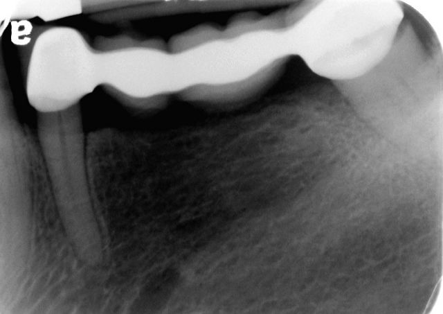
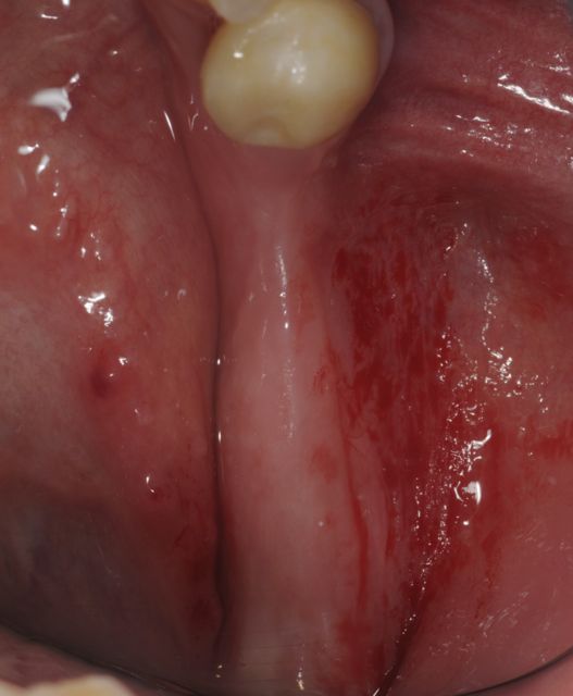
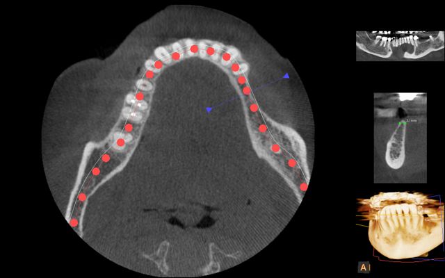
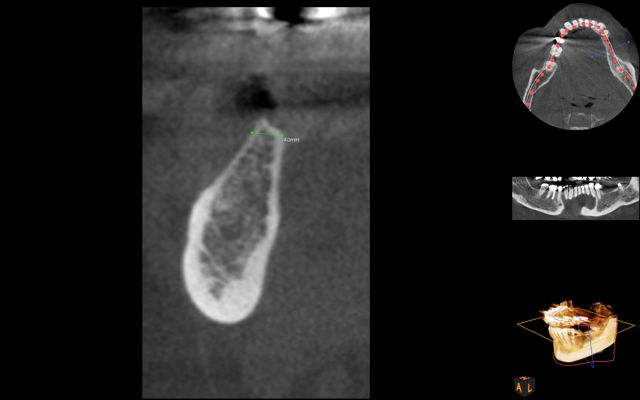
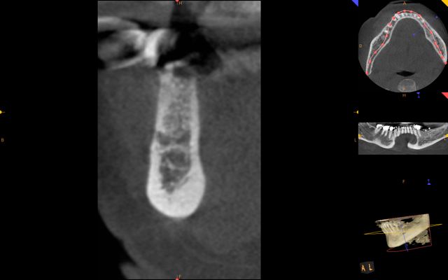
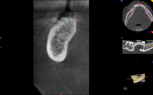
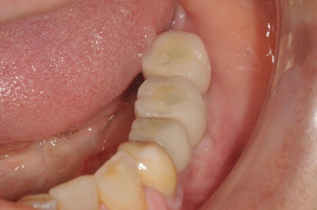
This patient presented with a failing bridge on her left lower jaw. The bridge had a long span and was loose due to recurrent decay. The bone where teeth were missing was quite thin due to the resorption process over time and had inadequate width for placement of dental implants. This was clearly demonstrated on CBCT (cone beam CT scan) . She did not want to resort to a removable partial denture and instead wanted to replace her missing teeth with dental implants. This required an initial bone grafting to restore the width deficiency of the bone and then replace her teeth with three dental implants.
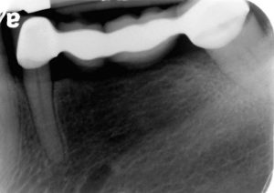
Failing dental bridge due to recurrent decay
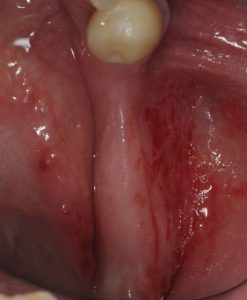
Missing teeth with bone loss inadequate for implant placement. Can be augmented with grafting.
Treatment:
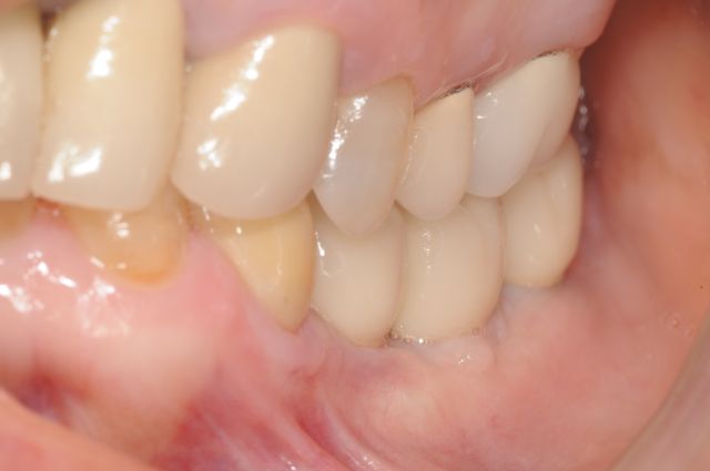
During the first procedure, Dr. Kazemi augmented the bone deficiency using an onlay bone graft technique under IV sedation. Following six months of healing, a new CBCT was taken to assess the augmented bone and a computer-assisted implant planning was completed. A surgical guide was fabricated for precise positioning of the dental implants according to her bite requirements. The implants were allowed to heal for 3 months. Finally, our team restorative dentist restored the implants with customized abutments and crowns.
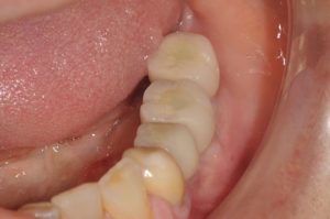
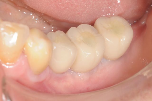
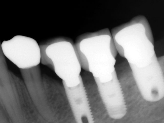
Bone was augmented with grafting. Then restored with three dental implants and single crowns.
Photo Gallery (Click on first photo to start):
Keys to achieving this result:
- Collaborative team approach between surgeon and restorative dentist
- Development of adequate bone foundation using onlay bone grafting technique
- Use of CBCT for 3-D planning and implant positioning.
- Precision placement of dental implants
- A surgical guide for implant placement based on crown positioning (prosthetic-based implant placement)
- Custom abutments and anatomically fabricated crowns with precision fit
