Bone Graft To
Increase Height
Schedule an appointment
About Bone Height Grafting
Sufficient bone height is critical for implant support and long-term success. Short implants have increased risk of failure, specifically in the back teeth where the forces of chewing are the most.
Loss of bone height may be caused by:
- Periodontal (gum) disease and gradual bone loss
- Trauma
- Denture use (partial or full dentures)
- Extraction of teeth without site bone grafting
- Downward shifting of the sinus floor following extraction of upper back teeth (first and second molars and occasionally second premolars)
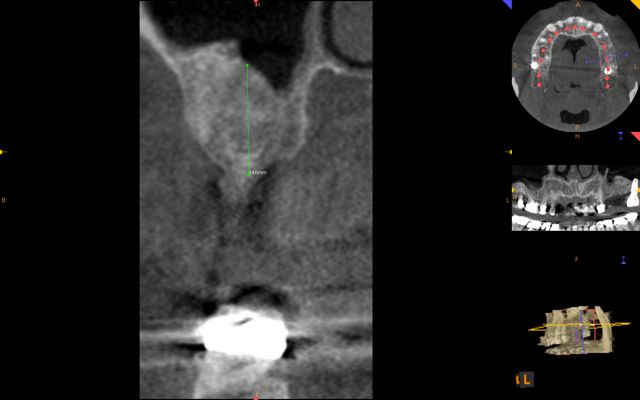
The amount of available bone is determined by clinical exam and a cone-beam CT scan. The planned site for the implants are then examined for the quantity and the position of the ridge and the proximity of vital structures such as the sinus and nerve canals.
Sites with deficient height of bone should be grafted for the following reasons:
- Selection of implants with proper length
- Improved positioning of the implant and alignment with the adjacent teeth
- Improved aesthetics
- Improved access for hygiene
- Prevention damage from high forces when implants are placed too deep
There are several techniques available for augmentation of bone in the vertical dimension based on location, degree of bone loss, and amount of bone augmentation needed:
Treatment is generally provided in two stages. In the first stage, bone grafting is done to correct deficiency in height, width, or both. Following 6 months of healing, dental implants are placed and allowed to heal. Finally the implants are restored with the final teeth.
Dr. H. Ryan Kazemi is one of the most experienced and innovative surgeons in the field of bone regeneration and grafting and has performed grafting procedures in thousands of patients.
Sinus Lift Bone Grafting
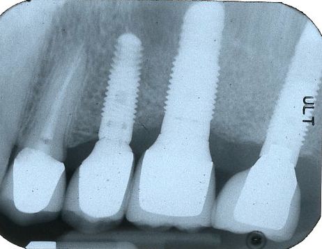
 Sinus lift bone grafting is indicated when the bone height is inadequate for implants in the back of the upper jaw due to proximity of the maxillary sinus. This occurs in the area of upper second premolars, first molars, and second molars. Following tooth loss, the maxillary sinus migrates downward due to local resorption and remodeling pattern. The migration of the sinus floor results in bone loss which often becomes inadequate for implant support.
How is the procedure performed?
Depending on amount of desired bone augmentation, a sinus lift procedure may be performed in 2 ways:
- External approach through a small window on the outer side of the sinus wall
- Internal approach through the site where the implant is placed (osseodensification technique)
The sinus membrane is gently lifted creating a small pocket where the bone graft material is inserted. Sinus lift procedure can be performed at same time as the implant placement if there is at least 5 mm of existing native bone height. Otherwise, it is performed in two stages where the bone augmentation is completed and allowed to heal for 6 months before placement of dental implants.
What materials are used for grafting?
The optimal bone material is combination of patient’s own bone and mineralized freeze dried bone available from bone bank. Small amount of bone can be obtained from the back of the lower jaw bone near the site of the wisdom teeth. In smaller bone height defects, bone bank by itself maybe used with great results. In-addition guided tissue regeneration membranes and Platelet-Rich Growth Factor are used to promote healing.
Is it safe?
The sinus lift procedure is a minimally invasive procedure and is quite safe. It is a safe technique with predictable results. Infection is the only potential complication which can be prevented through good post-operative oral hygiene and use of antibiotics. The procedure requires only a small pocket where the bone graft is placed, hence it does not alter the sinus function in anyway.
About the recovery:
Patients will experience some discomfort well managed by analgesics and swelling which resolves mostly within a week. Patients can return to work or school in 1-2 days.
See ‘Why Sinus Lift Bone Grafting is Not All That Big Of a Surgery’
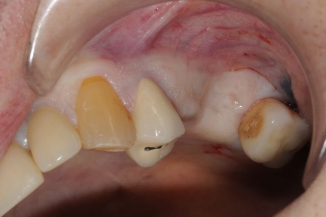
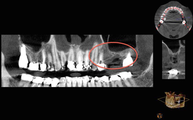
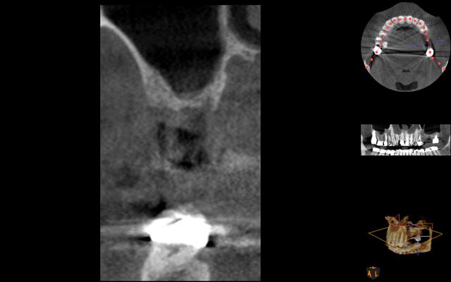
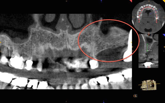
Patient example:
Ridge Bone Grafting
In some circumstances, the bone may be inadequate at the level of the ridge resulting in a vertical or height deficiency. This occurs commonly in teeth with chronic periodontal disease, trauma, or traumatic extraction with excessive bone removal.
Dental implants placed too deep at level of a deficient ridge have high risk of failure due to excessive load and poor access for hygiene. Hence, it is critical to augment the bone deficiency and improve the level of the ridge before placement of the implants.
There are two types of bone grafting techniques for improving ridge deficiencies:
Onlay Bone Graft:
Onlay bone grafting is indicated where there is minor to moderate bone deficiency at the top of the ridge resulting in inadequate height for implant placement. The techniques involves harvesting of bone from patient’s own mouth, commonly from where the wisdom teeth are, and then placed directly on top of the deficient ridge. The bone graft is then fixated and allowed to heal for 4-6 months.
The bone graft heals and becomes part of the jaw bone. Now the implants can be placed at ideal ridge level and restored to normal function.
Ti-Mesh with rhBMP2:
This technique involves use of a titanium membrane with bone graft material combined with bone morphogenic protein (rhBMP2). The titanium membrane holds the bone graft material in place and supports it during its healing phase. Typically, 6-8 months later, the titanium mesh can be removed and dental implants placed.
Interpositional bone graft:
A third technique for correction of vertical deficiency is known as interpositional bone graft. With this technique, the deficient ridge is surgically repositioned to the desired level and then graft is placed in the space between the ridge and the rest of the jaw bone. The segments are held with a fixation plate and allowed to heal for 6 months. At this time, the plate is removed and the dental implants are placed.
Graft Large Bone Defects
In circumstances where there are very large vertical bony defects, a distraction osteogenesis along with other ridge development grafting techniques may be recommended.
Distraction osteogenesis allows bone generation by gradually “stretching” the existing ridge using a unique device. As much as 7-10 mm of bone can be created in a week as the device is continually activated. Following a 3 month healing, the site is ready for either implant placement or further bone grating as indicated.
See a distraction osteogenesis case report on a patient with missing bone (**Note- Contains Surgical Photos**)
