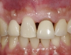
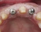
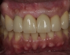
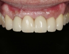
How Does Bone get thin?
Thinning of bone surrounding teeth can be caused by:
- Normal remodeling and shrinkage of bone that occurs following tooth extraction– this is particularly common in the upper front teeth (smile zone) but can occur with any tooth.
- Existing bone loss due to periodontal disease
- Trauma
- Difficult surgical extraction of a tooth that required bone removal
- Teeth sites that were not grafted at the time extraction
Loss of Bone Width will Result in:
- Difficulty placing a dental implant
- Loss of gum tissue
- Poor aesthetics
- Difficult cleaning
- Poor support for any type of tooth replacement (implant crown, conventional bridge, denture, etc.)
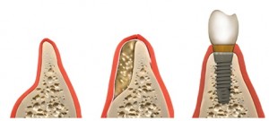
Horizontal Bone Grafting:
Horizontal bone grafting increases the bone width and allows for the proper positioning of an implant. Augmentation can be done either by using an onlay bone against the existing ridge or an expansion technique to increase it width.
Types of Horizontal Bone Augmentation:
- Onlay bone graft: The bone obtained from another area is laid on top of the existing bone and then stabilized using fixation screws. Calcified granules may be added to fill in smaller defects and then completely covered with a special protective membrane. The gum tissue is sutured and the bone is allowed to heal.
- Bone expansion procedure: A thin ridge may be expanded to increase its width. The space created is packed with some bone material to maintain its form and dimension.
- Ti-Mesh with rhBMP2: A titanium mesh is formed and stabilized with screws. The bone graft material is mixed with rhBMP2, a high concentrate of bone morphogenic protein, and placed within the mesh. Following 6-8 months of healing, implants may be placed.
Where Does the Graft Come From?
- Bone granules or particulates: These are calcified granules of bone, prepacked in sterile bottles, used routinely for bone regeneration. This type of bone graft material may come either from bovine bone, human bone, or synthetic in source. They are very safe and do not have any disease transmission potential. The material is treated so that only the calcified matrix remains with none of the proteins or other cellular components.
- Regenerative bone materials: There is great technology for developing bone in various regions using bone morphogenic protein. This allows for the regeneration of bone in various forms and amounts when combined with other forms of calcified bone tissue.
- Patient’s own bone:
- Bone from the ramus (back of the lower jaw bone where the wisdom teeth were): This is a very common source of bone for building width. It is relatively easy to obtain and is essentially similar to an impacted wisdom tooth removal procedure. Because this is the patient’s own bone, it is safe and carries no chances of disease transmission or rejection. It does not cause any cosmetic problems and it heals quite well.
- Bone from chin: Bone may be obtained from the chin area through a small incision in the gum tissue area. This does not cause any cosmetic deficiency or changes in the shape of the chin.
- Bone from hip: When a larger amount of bone is needed for significant defects, bone from the hip is often used. Bone is obtained from the inside bone of the hip and is not cosmetically visible. It is performed in a hospital under general anesthesia. The incision is very small and heals with minimal scarring. In females, the incision is carefully positioned within the bikini line.
Healing phase:
Bone grafts typically heal in four to six months, forming new bone identical to the shape of the transplanted bone. Implants can then be placed securely in this bone. It usually takes another two to five months for the bone to heal around the implants. Implants placed in these grafts generally have the same success rates as implants placed in non-grafted bone.
